News Story
Capstone 2010: Devices for Diagnostics, Improved Treatments, and Personal Health
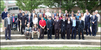
The Class of 2010 with faculty, mentors and staff.
Capstone, a two-part course taken in the fall and spring semesters of senior year, is one of the most important parts of the Clark School's engineering program. In it, teams of students utilize what they have learned throughout their undergraduate studies to create their own engineering designs from concept to product.
Mrs. Susan Fischell is the creator and sponsor of our annual Capstone Design Awards. In this competition, launched in 2009, the top three project teams as selected by a panel of judges win monetary prizes donated by Mrs. Fischell and are invited to present their work to the public at the Fischell Festival. The teams may also have the opportunity to have their inventions put on track for development at the Robert E. Fischell Institute for Biomedical Devices. Winners of the 2010 Capstone Awards are noted in the project descriptions below.
2010 Capstone II Teams and Projects
ThorLabs' QUANTA: Portable Quantification Imaging System
Jonathan Ament, Hsiang Lan Chan, Daniel Fam, Steven Graff, and Ziad Mahmassani
Faculty Advisor: Assistant Professor Yu Chen
Mentor: Dr. Suraj S. Venna, Washington Hospital Center
 Skin cancer is the is most common form of cancer diagnosed in the United States. Dermatologists and other doctors use both visual examination and biopsies to determine whether a skin lesion is cancerous, but a biopsy—taking a tissue sample by cutting away a small part of it—is required for a firm diagnosis. Since visual examinations are not quantitative and biopsies can be painful, the ThorLabs team worked on a new medical device called QUANTA, which can be used to provide skin cancer diagnoses using non-invasive laminar optical tomography. QUANTA applies light from a laser diode to the suspect area of skin, and senses the reflective differences between benign nevi (birthmarks, moles) and malignant or precancerous lesions. The device is designed to be portable and easy enough to use in any doctor's office. (Pictured: The QUANTA prototype, case opened to show its components.)
D.E.W.C.: Diagnostic Eletrophysiological Wireless Catheter
Joseph Davis, Farnoosh Farahi, Amanda Gravenhorst, Mads Matthiesen, and Tiffany Westo.
Faculty Advisor: Assistant Professor Ian White
Mentors: Dr. Amit Shah and Dr. Manish Shah, Washington Hospital Center
Second Place, 2010 Capstone Design Awards
 Arrhythmia, a condition in which the heart experiences abnormal contractions, is often treated with a surgical procedure called a catheter ablation. Two small, flexible tubes—catheters—are inserted into the chest. One, the guide catheter, is used by the surgeon to visually navigate through the patient's heart. Radiofrequency energy, transmitted through the second, is used to destroy (ablate) the tiny amounts of heart muscle identified as the origin of the problem, allowing the heart to beat normally again. Currently, the diagnostic information the surgeon receives from the guide catheter is processed by a junction box tethered to the device. This complex system of controls and wires requires intensive setup and is difficult to use. The D.E.W.C. team designed a guide catheter that transmits the same information wirelessly. The signal is amplified, digitized, and sent to a data acquisition system that provides the surgeon with a real-time display. The new device could reduce human error, the surgery time by as much as 45 minutes, and the cost of the procedure by $1000. (Pictured: The team demonstrates how D.E.W.C. transmits and processes signals.)
The Samet Group PocketDoc: Emergency Monitoring System
Omar Bekdash, Dipankar Dutta, James Pan, James Reilman, and Dennis Truong
Faculty Advisor: Professor Art Johnson
Mentor: Dr. Ron Samet, UMB School of Medicine
Third Place, 2010 Capstone Design Awards
 The PocketDoc is a portable device that supplements first response procedures by transmitting SpO2 (saturation of peripheral oxygen in the blood) and heart rate information to EMTs before or when they arrive on the scene of a medical emergency, or to doctors before a patient reaches the hospital, saving time and lives in the process. The device, which is designed to be easy to use by people without medical training, can display this quantitative information in real-time on a smartphone by connecting to its serial port, then transmit the same data wirelessly to 911 dispatchers or medical personnel. It is also capable of storing a patient's medical history. The Samet Group envisions the device becoming as common as emergency defibrillators in high-traffic locations such as malls, airports, and military bases, and schools. The device could be offered as a "plug and play" product for consumer use, as well as in a professional version for more advanced data logging. (Pictured: the prototype PocketDoc.)
GluCoach: Smartphone Interfacing Glucose Meter and Application (SIGMA) for Diabetes Management
Omar Ayyub, Andrew DeMaio, Katie Farhang, Nii Mante, and William Richbourg
Faculty Advisor: Professor Yang Tao
Mentor: Dr. Michelle Magee, Washington Hospital Center
TIE, First Place, 2010 Capstone Design Awards
 The GluCoach-SIGMA team used a smartphone and the Android operating system's software development kit to create a go-everywhere, easy-to-use device that enables diabetics to track their insulin, blood sugar, meals, exercise and more. Managing diabetes requires diabetics to stick to routines, plan meals, check their glucose levels, and administer insulin. The tracking of all of this information sometimes becomes burdensome, resulting in forgetfulness or mistakes. Noting that people almost never forget their cell phones, the GluCoach team designed a glucose meter capable of transmitting readings over Bluetooth to a smartphone installed with a suite of software designed to analyze the data, calculate insulin requirements, and help a person maintain a healthy lifestyle by keeping all the information they need at their fingertips. (Pictured: GluCoach running on a team member's smartphone.)
CAPRA: Capnographic Respiratory Analysis
Jennifer Lei, Allon Meizlik, Hector Niera, Charlie Sun, and Jessica Tsaoi
Faculty Advisor: Associate Professor Hubert Montas
Mentor: Dr. Amit Shah, Washington Hospital Center
 Dyspnea (difficulty breathing or shortness of breath) is a symptom of a variety of cardiovascular and respiratory conditions, many of them serious or life-threatening. It is one of the top five reasons for emergency room visits. Because dyspnea is such a common symptom, patients may experience a delay in the diagnosis of their underlying problem, or could receive inappropriate treatment due to a misdiagnosis. The CAPRA team designed a device that can be fitted into existing ventilator equipment used by hospitals and EMTs that is capable of differentiating whether a patient's dyspnea is caused by asthma, COPD, or pulmonary edema. It uses a combination of capnography, the continuous analysis of the concentration of carbon dioxide exhaled, and signal processing to amplify, transform and analyze signal data picked up from a sensor. The results are displayed as visual waveforms, which take on distinct, easy-to-interpret shapes that are indicative of whether the cause is an obstructive disease. (Pictured: The CAPRA prototype. The sensor at the end of the blue cable fits inside standard oxygen masks.)
COSMA: Collapsible Shape Memory Alloy Stent for Biliary Drainage
Jessica Bermudez, Dominique Franson, Elizabeth Kim, Megan Kuhn, and Pratiksha Thakore
Faculty Advisor: Associate Professor Elias Balaras
Mentors: Dr. Filip Banovac and Dr. Kevin Cleary, Georgetown Medical School; Dr. Robert E. Fischell; and Dr. Teng Li and Dr. Guangming Zhang, Department of Mechanical Engineering
 In the human body, the biliary tract is a network of ducts that transport bile from the liver through the common bile duct and to the small intestine, where it aids in digestion. If the biliary tract becomes obstructed (for example, by a tumor, gallstones, or inflammation), doctors may insert a thin, flexible tube called a catheter in order to drain excess bile. Multiple procedures may be required to insert, adjust, and remove the catheter, which increases cost, patient discomfort, and the risk of infection. Team COSMA designed a biliary catheter with a built-in stent—a tiny metal scaffold—made out of a shape memory alloy called nitinol. Initially, the catheter is very narrow, allowing doctors to insert it by making only a small incision. Once in place, voltage is applied to the catheter. When it reaches the portion containing the stent, the stent reacts by "unfolding," making the catheter wider and pushing open the blocked area of the biliary tract. When the catheter is no longer needed, heat is applied, which returns the stent to its collapsed state and the catheter to its normal diameter. Doctors are then able to remove the catheter through the same small incision site. The new product has the potential to make the procedure safer, more cost-effective, and less painful. (Pictured: Left: The catheter. Right: A large-scale model of the stent inside the catheter in its expanded state.)
Magnetic Particle Steering by Active Control of Magnetic Fields for the Purpose of Drug Targeting
Erik Li, John Lin, Chetan Parrija, Michael Tsai, and Jeffery Zhang
Faculty Advisors: Associate Professor Benjamin Shapiro and Assistant Professor Silvia Muro
TIE, First Place, 2010 Capstone Design Awards
 Many patients receiving chemotherapy for cancer become ill or die because of its side effects, a problem which has led doctors and scientists to explore how to accomplish more with fewer treatments. Magnetic drug delivery, in which magnetic fields are used to guide therapeutic magnetizable nanoparticles to specific regions of disease, is one possible solution. The Magnetic Particle Steering team worked on ways to improve the delivery of drug-carrying magnetic nanoparticles to deep tissue tumor sites. They developed an algorithm to model and predict the nanoparticles' behavior, a high spatial resolution imaging system capable of tracking them en route, and a biomimetic synthetic vascular system that simulates blood flow in the human body, which they used to test their ability to guide their nanoparticles through specific channels. The team demonstrated that their improved delivery system can result in up to four times more therapeutics reaching the site of tumor per treatment, which translates into less chemotherapy and fewer side effects for the patient. (Pictured: A still form a video of nanoparticles flowing through the team's biomimetic synthetic vascular system.)
Good Luck, Seniors!
We wish all of our seniors the best! We're sure innovative engineers like these will succeed wherever they go.
Thank you!
Our seniors would like to thank their professors and mentors (listed with their Capstone teams above); lab staff including Melvin Hill, Ali Jamshidi, and Gary Seibel for guidance, time, and labor; administrative staff members for help with financial support and purchasing; Professor and Fischell Department of Bioengineering Chair William Bentley; and friends in outside academia and industry for the advice and supplies they donated that helped these projects succeed.
The Fischell Department of Bioengineering would like to thank Mrs. Susan Fischell for her sponsorship of the Capstone Awards, and our panel of judges: Professor Leigh Abts (College of Education and affiliate faculty, BioE), Brian Lipford (Partner and VP of Strategic Initiatives, Key Technologies Inc.), and Dr. Jafar Vossoughi (President, Biomed Research Foundation and affiliate faculty, BioE).
Published May 18, 2010
Related Stories
Stories / May 17, 2023
24 Teams Present at BIOE Capstone Competition Including First...

Stories / September 12, 2022
UMD Bioengineering Advances in Rankings

Stories / May 9, 2022
BIOE Capstone Competition Highlights 24 Novel Projects

Stories / May 21, 2020
BIOE Capstone Class Virtually Presents 24 Novel Projects

Stories / May 17, 2019
BIOE Capstone Class Presents 20 Novel Projects
Stories / May 17, 2017
BIOE Capstone 2017: Projects Focus on Pediatric Care, Physical...
Stories / April 1, 2017
Getting Ahead of Alzheimer’s
Stories / June 28, 2016
BioE Capstone 2016: New Projects Focus on Improved Healthcare...
Stories / July 11, 2013
BioE Capstone 2013: New Products Focus on Diagnostics, Physical...
Stories / October 10, 2012
Partnership with UM School of Medicine Yields New Start-Up