News Story
BIOE Capstone 2018: Projects Focus on Point of Care Diagnostics, Sickle Cell Disease, Neonatal Care, and More

Showcasing the many ways bioengineers are working to improve human health, the Fischell Department of Bioengineering (BIOE) 2018 Senior Capstone class exhibited a total of 24 novel concepts during the Capstone II finale on May 7, 2018.
Projects ranged from a device for detection and monitoring of sepsis to a ventilator tubing support mechanism for use in the Neonatal Intensive Care Unit.
The 2018 Capstone Design Competition marked the biggest yet, as 116 students pitched their products to a panel of esteemed judges, as well as to BIOE faculty and fellow students. Created and sponsored by Mrs. Susan Fischell, wife of A. James Clark School benefactor Dr. Robert E. Fischell, the Capstone Design Competition has been a marquee event, capping off the end of the academic year.
This year’s panel of judges featured:
- The Fischell Department of Bioengineering Advisory Board
- University of Maryland, Baltimore collaborators
- The Bioengineering Graduate Student Society
2018 Capstone II Teams, Projects and Awards:
Team 1: Multi-channel Surface Electromyogram
Cailin Adair, Caroline Dong, Cameron Harner, Serenity Miller, and Malavika Shukla
Advisors: Dr. Yu Chen (BIOE Associate Professor), Dr. Yang Tao (BIOE Professor), Dr. Michael Dimyan (University of Maryland School of Medicine)
Electromyography (EMG) provides a method of studying muscle function by analyzing the electrical signals produced during muscular activation. It is used in the diagnosis of neuromuscular diseases and motor skill deficiencies like those seen in stroke patients. The most widely used method of EMG signal acquisition is needle EMG because of its accuracy. Even so, it is a painful, invasive and time-consuming procedure. Surface electromyography (sEMG) on the other hand, offers a way to noninvasively record muscle activity. However, the signal tends to be less accurate as a result of signal convolution. We propose an sEMG system, containing a multi-channel electrode array along with the required circuitry and data acquisition components for use in studying neuromuscular diseases. Specifically, we have constructed the hardware for signal acquisition and filtering, along with the software for graphic visualization. This system allows for the study of various muscles simultaneously as well as the identification of the electrode pairing which provides the clearest and most accurate signal. The application of this system has the potential to accelerate research related to the diagnosis and treatment of neuromuscular diseases.
Team 2: Point of Care Diagnostic Device for the Quantitative Monitoring of Chronic Cough
Jessica Baer, Lucia Fernandez, Andrew Kadish, Yousuf Naved, Farah Vejzagic
Advisors: Dr. Ian White (BIOE Associate Professor), Dr. Stella Hines (University of Maryland School of Medicine)
1ST PLACE

According to pulmonologists, chronic cough is very prevalent in the United States and can be caused by a range of illnesses, such as asthma, COPD, and various cancers. Additionally, many patients suffer from idiopathic chronic cough, for which a concrete cause has not yet been determined. Diagnostic tests currently available, however, fall short of characterizing cough. Instead, pulmonologists must use existing tests that measure different endpoints, such as airflow obstruction, to assess the severity of a disease, even though these diagnostic tests do not truly measure the symptom in question--chronic cough. Consequently, pulmonologists are unable to make accurate assessments about the efficacy of certain prescribed treatments. The purpose of this project is to invent a novel, cough monitoring device which will be capable of quantifying and monitoring chronic cough over long periods of time, thereby aiding pulmonologists in formulating an accurate assessment of cough severity and response to treatment. The proposed belt-like device is placed on the upper abdominal region and utilizes a piezoelectric sensor to detect changes in pressure caused by the contraction of the rib cage during coughing. The objective data collected by this device will then be transferred to doctors in real-time via a mobile application, giving them access to critical data pertaining to the cough of each individual patient. Patients will also be able
to input personal notes about their cough in the application. Preliminary testing of the prototype determined that coughing can be distinguished from other physiological events such as yawning and deep breathing. These results have been interpreted as “proof of concept” for our prototype design. This information provides means to further develop and test future designs of this device in the hope of producing a viable, sustainable product.
Team 3: Device for Accurate Placement of Chest Tube for the Treatment of Pneumothorax and Hemothorax
Andrew Broda, Havisha Garimella, Joseph Kim, Wesley Niswander, Troy Walker
Advisors: Dr. Joseph Rabin (University of Maryland School of Medicine), Dr. Lester Schultheis (BIOE Research Professor; Robert E. Fischell Institute for Biomedical Devices)
Pneumothorax and hemothorax are two conditions in which the air or blood, respectively, fill the pleural cavity, the space between the lungs and the chest wall. The presence of these fluids within the pleural cavity exerts enough pressure to cause a collapsed lung. In very mild cases, a patient’s pneumothorax or hemothorax may often resolve itself within a few weeks of bedrest and physician supervision. In more urgent cases, a chest tube is inserted between the ribs and fed to the dorsal side of the chest cavity to alleviate the pressure and extract the excess air or blood. Due to the immediate nature of the condition, a radiologist may not always be available for imaging assistance during the procedure. Our project aims to create a device that will help physicians accurately and efficiently insert the chest tube during thoracostomy procedures. Current technologies that utilize a camera are too expensive and require an additional operation, which would prove inefficient for this time-sensitive procedure. Other technologies that utilize an inserter, which is a cheaper and easier guiding system, still have associated complications rates, even with experienced physicians. Our team will work towards building a cheap, easy-to-use thoracic device which will lower existing complication rates due to insertional and positional errors. Issues of functionality and efficiency will be addressed to ensure our device can be used quickly, safely, and without a steep learning curve.
Team 4: Proximity-Activated Restraint Technology (Part) System
Aaron Cherian, Zachary Goddard, Natalie Livingston, Alex Pu, Michael Wong
Advisors: Dr. William E. Bentley (BIOE Professor; Robert E. Fischell Institute for Biomedical Devices), Dr. Jeffrey Hasday (University of Maryland School of Medicine)
Nearly 10% of ICU patients are restrained daily. Existing methods of restraining patients include wrist restraints, chemical restraints, and straitjackets, each one coming with its own complications, including injury, increased length of stay, and psychological damage. Thus, finding a more reliable, comfortable, and side effect-free solution to restraining patients in the ICU is an important need. In this project, we design a flexible restraint system called PART (Proximity Activated Restraint Technology). The system was designed around ease of use, comfort, mobility, reliability, and affordability. The goal was to create a standalone device that would allow for complete freedom of movement during the majority of use and would restrain only when triggered by an intended stimulus. The PART system is broken up into several components: a sensor, a lockable elbow brace, and a hand restraint glove. A highly specific inductive proximity sensor, unique to metals, detects areas of equipment or tubing covered in aluminum foil. Detection of a stimulus, specifically whether a patient is trying to tamper with medical equipment, switches the system from allowing full range of motion to completely restricting arm and hand motion. Upon detection of the foil, two locking mechanisms are engaged. The first locking mechanism is at the elbow, where a solenoid motor pushes a pawl into a gear, locking the elbow at a fulcrum and prohibiting any curling motion. The second locking mechanism of the device is the inflation of a pouch on the glove that prevents the patient from being able to grasp any tubing, wiring, or equipment that could lead to potential self-harm. The system will remain locked until a nurse or doctor resets the device. While this system is more expensive than the static restraints typically used, it will prevent additional injury to the patient and allow the patient to be released as soon as possible, saving the hospital thousands of dollars in additional days spent in the ICU.
Team 5: Surgical Instrument for Accessing and Preparing Bone Site for AgNovos Treatment
Justin Devos, Valerie Gupta, Allyson Meltzer, Ladeja Robinson
Advisors: Dr. John Stroncek (AgNovos Healthcare), Dr. John Fisher (BIOE Professor & Chair)
Osteoporosis is a condition characterized by a decrease in bone density, affecting over 44 million people in the United States. Osteoporosis is generally treated with a variety of medications including bisphosphonates, calcitonins, and hormone analogues/ receptors. Agnovos has been exploring a new, minimally invasive, procedure to treat osteoporosis through the development of an injectable biomaterial to replace and harden the site of diseased tissue inside of a bone - specifically in the hip. The tools in the current Agnovos kit used for the procedure are difficult to use because the surgeon has to handle multiple tools simultaneously. Also, the lack of a handle on the suction and irrigation tool makes it difficult for the surgeon to comfortably hold and maneuver it. An all-in-one device has been designed that combines the capabilities of the debrider and suction/irrigation tool to increase efficiency. The tool also incorporates an ergonomic handle to improve usability. Testing was performed on the new device to evaluate its debridement efficiency and suction capability compared to the current devices. The results show increased debridement efficiency with the new device and comparable suction capabilities between the new and current device. Overall, the new device would make the toolkit more effective and user-friendly, and subsequently increase the quality of life for many people with osteoporosis.
Team 6: Bedside Sputum Analysis System
Christopher Chester, Madelyn Golding, Alexander Lu, Cara Purdy, Alex Woo
Advisors: Dr. Jeffrey Hasday (University of Maryland School of Medicine); Dr. Gregg Duncan (BIOE Assistant Professor)
Physicians monitor sputum characteristics to inform decisions about starting, stopping and changing therapy for patients with acute and chronic lung diseases. Certain quantitative properties of sputum are altered in patients who have pulmonary diseases such as COPD, chronic asthma, and cystic fibrosis. Sputum changes are an important indicator of success or failure of treatment for respiratory infections. The physical characteristics of sputum are an important consideration when deciding whether patients are ready for removal of endotracheal tubes. However, current practice is limited to qualitative measures by nurses, respiratory therapists, and in the outpatient setting, patients themselves. Unfortunately, there is large inter- and intraobserver variability in the assessment of sputum, including elasticity and viscosity assessments.
These properties, if quantified by clinicians when treating patients, could provide key insight into improving the specificity of a diagnosis and treatment plan for each patient on a case-by-case basis. The goal of this project was to develop a user-friendly sputum collection system and a bedside or bench top device that will quickly, reproducibly, and objectively analyze viscosity and elasticity of the sputum sample. The quantitative and reproducible information about sputum physical characteristics provided by this system will improve decision-making about patient management as well as provide a discrete outcome for clinical studies of disease intervention. The modified sputum collection system was modeled using computer aided software and was designed to remain compatible with current hospital procedures for sputum collection if manufactured. A prototyped circuit design and code for the bench top analysis machine was developed and tested with a polyvinyl alcohol sputum mimic of varying concentrations. Quantitative values for viscosity and elasticity were obtained using the mimic, as well as an outputted stress vs. strain plot to further examine the sputum mimic characteristics.
Team 7: NIcool: Cooling Blanket for Premature Neonates
Evan Botterman, Amy Garcia-Vivas, Riley Keane, Vu Nguyen, Jimmy Vu
Advisors: Dr. Jocelyn Leung (University of Maryland School of Medicine); Dr. Hubert Montas (BIOE Associate Professor)
Hypoxic Ischemic Encephalopathy (HIE) is a condition suffered by newborns that is commonly treated with therapeutic hypothermia. Current treatments involve active or passive cooling mechanisms. A problem with these treatments involve overcooling the newborn, which can lead to further complications. Some hospitals do not contain the necessary equipment for therapeutic hypothermia. Thus, the newborns must be transported to a higher level NICU that is capable of providing appropriate treatment. Additionally, active cooling devices are bulky and inconvenient during transport. The goal of this project is to create a blanket that will cool the newborn to the target temperature range of 33.5-34.5°C, prevent overcooling, and be portable for transportation. The device’s design consists of a blanket with a compartment to hold a phase-change material (PCM) and temperature monitoring system. This mechanism is a combination of both active and passive cooling; a specific cooling mechanism is in place, but in order to prevent overcooling, a nurse has to remove the device. This is where the temperature monitoring system comes into play in which it provides an alert to the nurse when the temperature drops below the threshold. After testing, passive cooling at ambient temperature of 21°C took an average of 20 minutes to reach target temperature range. The 15°C and 1°C PCM took an average of 10.5 and 4.5 minutes, respectively. In conclusion, the 15°C PCM cooling effect is not as abrupt compared to the 1°C PCM, but is considerably quicker than ambient temperature. The 15°C would be sufficient enough for temperature reduction and avoid temperature shock to the newborn. Future works include investigating temperature sensor accuracy and precision, reduction of temperature sensors used, incorporate esophageal temperature probe, and automated blanket removal system.
Team 8: Implementing a High Flow and Purity Oxygen Concentrator for Low Resource Settings
Sarah Aryee, Naomi Feldman, Stephanie Hutchinson, Anthony Nam, Sarah Taylor
Advisors: Dr. Angela Jones (BIOE Senior Lecturer), Dr. Adnan Bhutta (University of Maryland Medical Center)
The product of interest for this capstone project is a low-cost oxygen concentrator that can be used in developing countries, specifically for pediatric patients. Oxygen concentrators are used when a patient cannot maintain their own breathing or oxygen levels. Most children who present severe respiratory distress are in the developing world, and 25-50% of these children die. According to Dr. Bhutta, current oxygen concentrators that are in use in third-world countries do not deliver a high enough flow rate or concentration of oxygen in order to be effective. Flow is important because when there are a lot of secretions in the airway, airflow becomes obstructed. The increased flow opens the obstruction which keep the lungs open. Current third-world devices only supply about 2 L/min, which is not nearly enough for proper mechanical ventilation. The proposed device would ideally deliver between 4 and 15 liters of oxygen per minute with an oxygen concentration of at least 60%. Additionally, the device should be able to be powered by a battery (for areas that do not have electricity), but be adaptable to electrical outlets. Using these power sources, the oxygen concentrator should be able to run for 8-12 hours at a time, as this is the length of a typical nursing shift. Some patients may require the use of the oxygen concentrator for days at a time, hence the need for a power source that can easily be switched out or changed in areas with unreliable electricity. Another important factor of the proposed device is maintaining a low cost. Current oxygen concentrators used in third-world countries cost about $300-$400, so the cost of the prototype built by the team must be comparable in price. Although the cost will be low, the materials used or the oxygen concentrator must still be safe and reliable with minimal leakage. Finally, the device should be portable or at least on wheels. As this capstone project is a continuation of a project from last year, the main focus will be to build a functional prototype that builds on the progress made by the previous team. The key elements of the proposed design that set it apart from current commercial oxygen concentrators and last year’s work are: 1) a cyclical flow of oxygen for increased purity, 2) a valve for zeolite depressurization, 3) the use of a feedback loop based on oxygen for valve control, and 4) the use of only one molecular sieve bed. As a result of these improvements, this oxygen concentrator should meet the necessary flow, concentration, cost, and transportation elements necessary for success in developing countries.
Team 9: Point of Care Ultrasound-Stethoscope Device for General Diagnostic Applications
Brandon Chu, Allen Luk, Riddhi Gopal, Joe Puthumana, Devin Wright
Advisors: Dr. Jean Jeudy Jr. (University of Maryland, Baltimore), Dr. Li-Qun Zhang (BIOE and University of Maryland School of Medicine Professor)
2ND PLACE

Although some may claim that the stethoscope has long since outlived its usefulness, the adoption of stethoscope capabilities into an ultrasound, which has the ability to share data, may be able to prevent misdiagnosis and aid clinicians in identifying heart and lung related disorders. Traditionally, stethoscopes and ultrasounds are used independently from one another. This creates a time discrepancy where the live visual data obtained from the ultrasound is not in sync with the auditory data obtained from the stethoscope. In addition, there is also a matter of convenience where clinicians have to switch devices. The solution to these issues would involve a stethoscope-ultrasound combination that retains the functionality of both individual devices. This would allow the collection of both auditory and visual data simultaneously in order to provide a more accurate diagnosis. In order to physically attach the stethoscope and ultrasound in the prototype, a clip was 3D printed, allowing both probes to be removed, if necessary. The synchronization and display of outputs from the stethoscope and ultrasound, was done via MATLAB software, showing the user both sets of data at the same time. In the future, the clip could be modified to be more ergonomic for the user, different stethoscope and ultrasound probes could be used to provide better signal, and real time synchronization would be performed to provide more clinical utility. Furthermore, the ability to convert outputted data to an electronic version would be beneficial in generating and updating electronic records of tests performed on a patient for reference at a later time and objective analysis by multiple professionals.
Team 10: Temperature-Controlled Water Pad to Remedy the Effects of Hypoxic-Ischemic Encephalopathy in Newborn Infants
Marisa Adelman, Dylan Donley, Audrey Heffner, Lily Motabar, Lisa Singh
Advisors: Dr. Keith Herold (BIOE Professor), Dr. Jocelyn Leung (University of Maryland School of Medicine)

Hypoxic-ischemic encephalopathy (HIE) is a brain injury caused by oxygen deprivation to the brain in newborn babies. The preferred method used by doctors to combat HIE is to induce mild hypothermia (33.5-34.5 degrees Celsius body temperature) within 6 hours of diagnosis in order to preserve neuronal structure and function. 1 The current process used to induce mild hypothermia is passive cooling, in which the newborn infants are exposed to the environment and their body temperature is monitored. If the newborn becomes too cold, they are wrapped in a blanket. If the newborn becomes too warm, they are cooled down using ice packs and by removing blankets. Our project proposes utilizing a self-regulated temperature-controlled water pad to maintain the desired body temperature range without the need for constant monitoring. Peltier chips are used to control the water temperature flowing through the water pad that will affect the infant’s body temperature. A heat exchanger transfers the Peltier temperatures to the water. After developing a prototype of our product, we found that the Peltier chips were able to adjust water temperature within a range of 18 degrees Celsius to 34 degrees Celsius. This large range of water temperature would be able to easily affect the body temperature of the infant. Future work includes further development of the feedback loop to use the skin temperature of the baby to adjust the temperature of the Peltier device, and additional tests on the Peltier chips to improve the performance of the cooling curves.
Team 11: AutoNutriFlow: Automated Feeding Pump
Negin Haghighi Ara, Boris Deogracias, Griffin Mess, Jane Rouse
Advisors: Dr. Li-Qun Zhang (BIOE and University of Maryland School of Medicine Professor), Dr. Avelino Verceles (University of Maryland School of Medicine)
Muscle atrophy is the wasting or loss of muscle tissue via lack of protein and muscle degeneration in the body and can affect up to 90% of critically ill patients, mainly in ICU units. The decrease in muscle mass can significantly impact the rehabilitation time and recovery after hospitalization. The primary reasons causing muscle atrophy are insufficient protein intake and reduced muscle use. While there are many devices on the market that focus on increasing physical exercise and muscle movement in patients, there have not been significant improvements on the efficiency of gastroenteric feeding tubes which are the primary method of nutrition intake in ICU patients. Therefore, Group 23’s project focuses on preventing muscle atrophy and ensuring proper nutrition intake by identifying and correcting the existing gastroenteric feeding tube inefficiencies. One of the main reasons of insufficient caloric intake is the errors and inaccuracies that may arise when determining the correct flow rate of the nutrient dense solution. Currently, feeding tube flow rates are determined and adjusted manually by nurses, using a feeding schedule and pump flow rate chart that takes into account different variable such as patient’s daily caloric needs and gap times in between feeding intervals. This manual adjustment process allows for human error and miscalculation of the correct flow rate of the nutrients which may ultimately result in muscle atrophy. Moreover, overfilling and built-up pressure due to excess residual volume within a patient’s stomach is another problem with current feeding tubes which can cause complications such as vomiting or bloating. Recognizing this, Group 23 designed and developed an automated feeding pump that automatically calculates and adjusts the flow rate of the nutritional solution based on multiple variables such as outputs from a pressure sensor attached to the distal end of the feeding tube to measure the residual volume within the stomach, and a timer that accounts for time the patient would have been disconnected from the feeding tube system for reasons such as surgery and therapy. In addition, the system is designed to calculate the basal flow rate based on patient’s personal medical information such as physical characteristics, and nutritionist recommended caloric intake. The automation and the use of sensors in the feeding tube will ensure that the proper amount of calories is getting fed into the patient per day and therefore resulting in a lower percentage and chance of muscle atrophy in patients.
Team 12: Magnetically Guided Nanogastric Feeding Tube
Noam Gannot, Alston Kau, David Nguyen, Tyrus Vong
Advisors: Dr. Javier Atencia-Fernandez (BIOE Research Assistant Professor), Dr. Steven Tropello (CoapTech), Elisabeth Goldwasser (CoapTech)
Nasogastric tube (NGT) insertion is a common procedure performed by residents and nursing staff to access the stomach of patients in intensive care units (ICUs). This is a seemingly simple procedure that is usually performed at a patient’s bedside. The most common method of insertion is to insert it blindly and then confirm placement of the tube in the stomach using X – ray. Complications that arise from improper placement of the tube are well known and, in some cases, may be life threatening. Furthermore, this method is difficult to perform in non-cooperative patients.
Our Capstone team, along with CoapTech, propose a solution to ensure proper placement of an NGT and a method that will be applicable to those patients who have difficulty undergoing the normal procedure. This will be done using a novel patented technology that combines magnetic guidance and ultrasound visualization. Strong rare- earth magnets will be used to guide the device placement inside the body and real-time ultrasound will provide visual feedback to the clinician. This technology, termed Coaptive Ultrasound TM has been used to insert gastrostomy tubes and has been validated in cadavers and live canines. Coaptive Ultrasound TM will be utilized to more efficiently and safely insert NGTs and offer reduced healthcare costs, improve safety, quality, and patient experience.
Proof of concept of the use of Coaptive Ultrasound ™ for the insertion of NGTs was obtained. Attraction between an external magnet and an internal magnetic tip was established at a distance of 10cm. Furthermore, through cadaver testing, visibility of the magnetic tip was established. While further testing is still needed, the use of magnetic guidance and ultrasound visualization has great potential for an NGT application.
Team 13: Streamlined Mixing of Bone Cement for Osteoporosis Treatment
Monica Chu, Patrick Grandinetti, Manny Mastromanolis, Veda Ravishankar, Tiffany Wun
Advisors: Dr. Christopher Jewell (BIOE Associate Professor), Dr. Jonathan Shaul (AgNovos Healthcare)
BEST ABSTRACT

Osteoporosis is a systemic metabolic disease that results in abnormally porous bone, which in turn leads to decreased bone density and strength. The resulting bone is fragile and prone to frequent fractures. Approximately 200 million women worldwide are currently affected by osteoporosis, while 1.6 million suffer from a hip fracture every year (Osteoporosis Facts and Statistics, 2017). Current treatment options include bisphosphonate-based pharmaceuticals, which have low compliance rates among patients and take up to 6 to 12 months to take full effect (Harding, 2017). As a result, a new adjunctive treatment known as local osteo-enhancement procedure (LOEP) has emerged, involving the injection of a biocompatible material, commonly known as bone cement, into the femoral neck of osteoporotic hips in order to strengthen them. Although there are a number of functional devices currently available on the market for the mixing and delivery of bone cement, there exists substantial opportunity to improve the process. After thorough analysis of the current mixing system used by AgNovos Healthcare, we noticed a number of opportunities for improvement, specifically in overall intuitiveness, ergonomics, and interaction between kit components. As a result, our group aimed to advance the current mixing strategies by creating a streamlined instrument to mix bone cement that would be injected into the osseous void via a minimally invasive method. Through torque analysis and material loss testing with a 3D printed prototype, we determined that our device is able to reduce material loss by 30.3% and reduce torque by 40.83% . This demonstrates that a manufactured version of our device has the potential to reduce material waste and increase ergonomic efficiency of existing bone cement mixing which will improve LOEP for osteoporosis treatment. Looking ahead, our team plans to redesign our device so that it will allow manufacturing with a plastic injection mold to simplify the manufacturing process and reduce the manufacturing costs. Furthermore, we plan to repeat our verification testing with a more accurate prototype composed of materials better suited for a working prototype. After verification testing, we will perform validation testing to confirm that our device has met user needs. Finally, it is our team’s hope to complete the provisional patent process with UMD’s Office and Technology Commercialization, ultimately to secure any and all IP specific to this project.
Team 14: Continuous Heart Rate and Blood Pressure Monitor for Detection and Monitoring of Sepsis
Fade Aromolaran, Mairead Fahy, Kara Huie, Elene Nakas, Brooke Szczesny
Advisors: Dr. Adnan Bhutta (University of Maryland School of Medicine), Dr. Lan Ma (BIOE Lecturer)
While sepsis is a universally deadly condition, its mortality rate in the developing world is especially high. Because of a lack of funding for the necessary equipment and infrastructure for proper diagnosis and treatment of sepsis, the mortality rate of sepsis in third world country hospitals ranges up to 60%. To help lower this statistic and improve chances of survival of septic patients, we have developed a prototype of a wearable device that continuously monitors heart rate variability (HRV), a proven indicator of sepsis. Our prototype is able to amplify and filter voltage signals from a piezoelectric sensor, display the signal over a time scale using Matlab, and calculate heart rate. We were also able to make preliminary HRV calculations from our signal, although further optimization is needed to be performed for better accuracy. With further development, our device would be able to also monitor blood pressure variability (BPV) and use these two parameters to determine whether the patient is at risk of sepsis onset. Future work would also include the implementation of machine learning capabilities to determine whether the patient’s HRV and BPV align with that of a septic patient. Ultimately, we hope to accomplish the intended social impact of decreasing the mortality rate of sepsis in the developing world.
Team 15: Real Time Distance Monitoring to Refine Placement of Endotracheal Tubes
Urvashi Dayalan, Jacob Gross, Thomas Mathews, Michael Reilly, Jessica Yau
Advisors: Dr. Ed Eisenstein (BIOE Professor) Dr. Dr. Dayanand Bagdure (University of Maryland School of Medicine), Dr. Jason Custer (University of Maryland School of Medicine)
The proper placement of an endotracheal tube (ETT), a flexible plastic tube inserted into the airway of a patient during endotracheal tube intubation (ETI), facilitates the mechanical ventilation of a patient and maintains airway management during intensive care and procedures requiring anesthesia. ETTs are often prone to slippage, moving out of their optimal position in the trachea and preventing proper ventilation of the patient. Immediate and frequent monitoring of an ETT is of the utmost importance to maintain patient safety throughout the duration of for which a patient requires mechanical ventilation. Failure to properly insert an ETT or maintain the position of an ETT can lead to hypoxaemia, severe hypotension, oesophageal intubation, and cardiac arrest. Current techniques to confirm placement of an ETT within a patient include chest x-rays, laryngoscopy, and esophageal detectors. However, these methods are invasive, lead to frequent radiation exposure, and do not provide continuous readings. ETT visualization and monitoring is particularly important in intensive care unit (ICU) patients, as they are often required to be intubated for weeks to months. This prolonged need for an ETT and thus for ETT visualization can lead to many complications, such as increased risk of developing cancers and a number of other comorbidities due to the current method most used for visualizing ETT placement, chest radiography. Our goal is to modify the ETT to provide a sensory relay that will visualize the ETT within the trachea to provide real-time monitoring of the distance from the end of the ETT to the carina, thus maintaining proper placement of the ETT in ICU patients and preventing the need for frequent exposure to ionizing radiation.
Team 16: KABAE Tube: A Triple Lumen Device to Reduce Microaspiration-Related Mortality in Neonatal Intensive Care Units
Erica Brown, Katie Geary, Ajay Kurian, Adam Paley, Bryan Wand
Advisors: Dr. Kimberly Stroka (BIOE Assistant Professor), Dr. Sripriya Sundararajan (University of Maryland Medical Center)
3RD PLACE

Microaspiration is the unintentional entry of liquid from the stomach into the respiratory tract which can lead to serious medical issues including bacterial overgrowth, pneumonia, and mortality. Neonates are at increased risk of microaspiration due to common breathing issues such as chronic lung disease or respiratory distress syndrome, as well as a lack of developed airway and cough reflexes. Additionally, many premature neonates cannot tolerate food in the stomach and require jejunal or duodenal feeding. Neonates with respiratory issues that require a nasotracheal breathing apparatus often additionally require nasojejunal feeding. Due to limited space to insert tubes in these premature neonates, feeding and suctioning tubes are not able to be place simultaneously, and jejunal feeding tube slippage into stomach is not able to be identified until the tubes are checked or a microaspiration event occurs due to increased intragastric pressure and gastric fluid content. Simultaneous continuous feeding and intermittent suctioning would allow nurses to be alerted to tube misplacement, and reduce the likelihood of microaspiration events and therefore neonatal mortalities. The KABAE Tube is a triple-lumen feeding tube with feeding, suctioning, and venting capabilities that can be used concurrently with a breathing tube. The KABAE tube performs with equivalent efficacy to standard feeding tubes while remaining compatible with the NICU infrastructure.
Team 17: VitalsWrap: Integrative Blanket for Hypothermia Prevention of Premature Infants
Jacqueline Dempsey, Alana Gudelsky, Jacob McCright, Nicole Palumbo, Julia Ringel
Advisors: Dr. Rose Viscardi (University of Maryland School of Medicine), Dr. Steven Jay (BIOE Assistant Professor)
Hypothermia is an extremely prevalent and dangerous condition that affects more than 40% of all premature newborns directly following birth. Aside from mortality, neonatal hypothermia can contribute to other complications including morbidity from acidosis, delayed transition from fetal to newborn circulation, abnormal coagulation, infection, and respiratory distress syndrome. To combat this, premature newborns are wrapped immediately after birth in a heat-retaining polyethylene wrap. However, this wrap can be improved through the integration of a variety of non-invasive monitoring sensors including: electrocardiogram leads, a pulse oximeter, and temperature probes to give the physicians and nurses more quantitative information on the health of the premature newborn. Our project integrated these sensors and a sodium acetate warming solution into a polyethylene plastic wrap design to reduce 1) the time required to stabilize the premature infant immediately after birth; and 2) the incidence of hypothermia and associated morbidities in these critically ill infants. Results concluded that the VitalsWrap performed approximately 4.4 times faster than the commonly used, NeoWrap TM . With further testing and prototyping, this device could be used as the new standard of care in NICU resuscitation procedures, significantly improving the quality of care and chances of survival for premature infants.
Team 18: Prosthetic Integrated Bone-Anchored Hearing Aid
Carrie Baumgartner, Nidhi Gandhi, Timur Kamalitdinov, Connie Nguyen, Jesse Vo
Advisors: Dr. Giuliano Scarcelli (BIOE Assistant Professor), Dr. Diego Preciado (Children’s National Medical Center), Dr. Kevin Cleary (Children’s National Medical Center)
One in 10,000 children are born with microtia, a condition in which the external ear and/or ear canal are absent. However, these children tend to have functional inner ears. This can lead to problems with development of speech and hearing. In addition, the physical deformity of missing an ear can have devastating consequences on a child’s emotional and social development. Current solutions provide children with a bone conduction hearing aid (BAHA) and a separate ear prosthetic. BAHA devices are implanted in a child’s skull, and vibrate in response to sound waves. The vibrations are transmitted to the inner ear, which allows for hearing. However, these solutions are noticeable and can still leave a child feeling isolated. We developed a solution for these children by combining the two existing technologies: a moldable ear prosthetic integrated and a bone conduction hearing aid circuit. The circuit transmits sound to an amplifier via a microphone, from there the sound wave travels to a bone conduction transducer. This transducer connects to a titanium screw, which are often used to attach ear prosthetics to the skull. The screw vibrates in accordance to the transducer’s vibrations, allowing the noise to travel to the inner ear. To test the viability of the device, we utilized a sound booth to generate an audiogram, which shows the softest noise a patient can hear at different frequencies. Compared to market BAHA devices, our prosthetic did not achieve the same hearing thresholds. We expect a significant improvement in hearing performance with a more robust circuit.
Team 19: Freemoglobin: Removal of Free Hemoglobin
Christine Anderson, Metecan Erdi, Stephanie Hughes,Thomas Mumford, Sarah Staines
Advisors: Dr. Giuliano Scarcelli (BIOE Assistant Professor), Dr. Michael Mazzeffi (University of Maryland School of Medicine)
According to the American Red Cross each year 4.5 million people in the U.S. need lifesaving blood transfusions. Thirty-six thousand units of blood are transfused every day in the United States. Units of blood can be stored for up to forty-two days before they are destroyed and discarded. The longer blood bags are stored the higher potential for negative effects of the transfusion such as renal insufficiency, high blood pressure, and various other adverse effects of the blood. As blood bags age, potassium levels increase which in turn decreases cell membrane integrity leading to hemolysis. Hemolysis releases free hemoglobin into the blood which causes the negative effects. The current method of free hemoglobin removal is to wash the blood using several centrifugation steps which causes shear stress on the viable cells rendering them susceptible to hemolysis4. Blood washing is only done in severe cases for immunocompromised patients such as babies and the elderly. To solve this problem, a filtration system was created that allows stored blood to flow through a filter into a new blood bag before transfusion into the patient. The filter is comprised of a nitrocellulose membrane that is housed in a polycarbonate tubing to absorb free hemoglobin and other proteins from the blood. Nitrocellulose membranes are used in labs everyday for protein absorption. To test the efficacy of the nitrocellulose membrane both a Lowry and BCA assay was performed to quantify the protein concentration in the blood before and after flowing through the filter and at various flow rates. Along with this, cell counting using a hemocytometer was performed before and after filtration to ensure that the red blood cell count remained normal. It is expected to see a decrease in protein concentration after filtration which correlates with a decrease in free hemoglobin. The results of the BCA and Lowry assays indicate a decreasing trend in protein concentration as the exposure time to the nitrocellulose membrane increased. In future, the filtration system could be used as the new standard in the blood transfusion protocol.
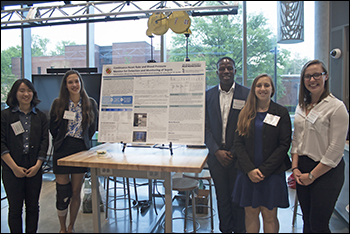
Team 20: Confronting Incompressible Torso Hemorrhage: Transesophageal Aortic Magnetic Occluder (TEAMO)
Turki Alahmadi, David DuBey, Kellie Holovac, Zoë Mote, Rohan Sivam
Advisors: Dr. Joseph Rabin (University of Maryland School of Medicine), Dr. Zhongjun (Jon) Wu (University of Maryland School of Medicine, BIOE Research Professor)
MPOWER AWARD
BEST POSTER AWARD

Approximately 500,000 people bleed to death every year in the U.S., and many of these deaths are a result of a specific type of trauma: incompressible abdominal hemorrhage. This type of trauma can only be treated by rushing the patient to a surgeon as quickly as possible, as no consistent field technology exists to stop major abdominal bleeding. As a result, patients often die before ever reaching an operating room. We have developed a medical device, the TransEsophageal Aortic Magnetic Occluder (TEAMO), in order to extend the life of patients in traumatic hemorrhage condition. TEAMO is a minimally invasive and easy-to-use device that occludes blood flow in the descending aorta. The device is composed of two parts. The first part is an esophageal tube with an attached internal magnet that is inserted through the mouth, and then into the esophagus to a precise location above the gastroesophageal junction. At this location the esophagus, aorta, and spine overlap. The second part of the device is an external electromagnet that is placed behind the patient’s spine. Once the electromagnet is activated, it will pull the internal magnetic device in the posterior direction and into the aorta, sandwiching it against the spine. This force will effectively clamp the aorta. This method of occlusion cuts off blood flow to the injured area and lower body, but it maintains flow to the brain, lungs, upper chest, and arms. To design this device, the force requirements of aortic occlusion was experimentally obtained. Using a resected segment of porcine descending aorta and a blood pump to model physiologically relevant flows and pressures, the required force to fully occlude the aorta was determined to be just under 10lbs. In addition, a cadaver of an average sized person was examined to obtain the approximate distance between the inside of the esophagus and the back of the spine, which is a constraint necessary on the design of the external electromagnet. With these design constraints and prototype, it will be possible to design a functioning device with a customized electromagnet in the future.
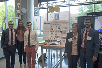
Team 21: Neovibe Device for Apnea of Prematurity
Nicholas Cha, Sukriti Ghosh, Geena Lingberg, Aafreen Parvez, Sonya Williams
Advisors: Dr. Rose Viscardi (University of Maryland Medical Center), Dr. Yang Tao (BIOE Professor)
STUDENTS’ CHOICE AWARD
Premature apnea is a pause in breathing longer than 20 seconds in infants. Short-term consequences of apnea in premature infants include oxygen desaturation and bradycardic episodes which, when prolonged, can lead to brain injury. Current treatment at the University of Maryland Medical Center (UMMC) involves tactile stimulation from a nurse. Once a nurse is alerted to an apneic episode, they must sanitize their hands, open the isolette, and provide the tactile stimulation, generally by rubbing the infant’s back. This process decreases response time, and compromises the sterile environment within the isolette, increasing potential for spread of infection in the neonatal intensive care unit (NICU). The novel NeoVibe device provides a standardized and sterile treatment for apnea of prematurity by using vibrational stimulation to end infant apneic episodes. The device consists of a grid of vibrational motors and is placed inside the back pocket of a SnuggleUp, a soft positional aid currently used for premature infants at the UMMC. Using Radio Frequency (RF) communication, the device is controlled via a wireless remote and eliminates the need to open the isolette, reducing the threat of infection while providing a quicker response to apneic episodes. The remote control includes three intensity settings for the vibration applied to the back that correspond to differences in pulse width modulation. This allows the nurse control over the intensity of the stimulation to prevent habituation to the device. This procedure standardizes the protocol in regards to treatment of premature infants experiencing apneic episodes in the NICU.
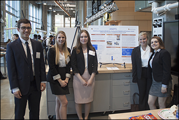
Team 22: Incentive Spirometry for Sickle Cell Patients
Joanne M. Chan, Nicholas S. Giroux, Andrew B. Lippe, Shivani U. Patel, Elizabeth A. Wilson
Advisors: Dr. Kevin Cleary (Children’s National Medical Center), Tyler Salvador (Children’s National Medical Center), Dr. Gregory Payne (BIOE Professor)
ADVISORY BOARD AWARD FOR TRANSLATIONAL DESIGN

Patients with sickle cell disease suffer from reduced oxygen-carrying capacity that results in abnormally low blood-oxygen content. To combat this and prevent the development of other pulmonary complications, a therapeutic lung distension technique, incentive spirometry (IS), is prescribed. Although IS is effective and inexpensive, research shows underutilization of this technique in adolescent patients likely because proper and regular use of IS can be difficult to encourage without clinician supervision. This project aims to engineer a digital incentive spirometer that promotes the regular use of IS by patients aged 7-19 and collects longitudinal data accessible to their clinicians. The spirometer mechanism consists of a turbine in tandem with an infrared LED to measure air flow and calculate peak expiratory flow, forced expiratory volume in one second, and forced vital capacity. The spirometer connects to a computer via a USB cable and works with an interactive game that adolescent patients will play while completing their lung exercises. Users are prompted to inhale and exhale to play the game while also providing data for downstream analysis. The current prototype was successfully calibrated against a known volume of air, and the observed flow measurements fall within gold-standard references ranges provided by the CDC. Integration of data collection with the interactive game mechanic, created on Stencyl, is ongoing.
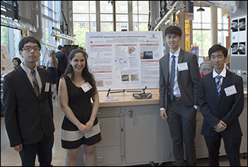
Team 23: Ventilator Tubing Support Mechanism for Use in NICU
Emma Dobry, Samuel Han, Christopher Mikus, Sri Krishna
Dr. Rose Viscardi (University of Maryland Medical Center), Dr. Helim Aranda-Espinoza (BIOE Associate Professor)
Premature babies in the Neonatal Intensive Care Unit often have difficulty breathing because their lungs aren’t fully developed. In addition, complications of labor and delivery, birth defects, and infections all can result in the infant having breathing problems and requiring the assistance of a ventilator. Mechanical ventilators push warm air and oxygen into the infant’s lungs through an endotracheal tube placed into the infant’s windpipe through the mouth or nose. A variety of methods are implemented in order to ensure endotracheal tubing remains fixed within the infant’s windpipe. However, with accidental extubation being a common outcome across various fixation approaches, endotracheal tube slippage, tube malposition, and the need for re-intubation can cause adverse effects such as mortality, perioral skin trauma, tube re-taping, and developmental complications. A current method of securing tracheal tubing is by supporting the two ventilator tubes, one pumping warm air and oxygen into the lungs and another pumping air out of the lungs, in order to stabilize the attached endotracheal tube. A product in the market commonly used in NICU’s across the nation is Poltex’s Angel Frame. Secured under the infant’s mattress, this product is composed of a wheel with grooves for securing the ventilator tubes. The wheel can be rotated and translated vertically in order to adjust the position of the ventilator tubing before fixation. This simplistic design effectively secures the endotracheal tube in the infant’s windpipe by fixing the ventilator tubes, which in turn secures and holds up the attached endotracheal tube in the infant. However, the rigidity of this design fails to address the more complex problems of ensuring the tracheal tubing remains in position with the infant lying in variable orientations in three-dimensional space while also moving unpredictably. Therefore, a novel ventilator tube support system is necessary that will improve upon the Angel Frame design by having the flexibility to accommodate for variable orientation of the infant within an incubator and the ability to adjust with the baby’s movements. This new design will be of great help to clinicians in the NICU by reducing the frequency of accidental extubation of infants that need help breathing. Our design includes a telescoping arm with a hanging wire to allow tubing to move up and down with baby movement and a tracking system using Kinect to sense when to translate side to side on the frame. We designed the frame to match our design and are using PEX-b to construct most of the components. At time of print no testing has been completed due to prototype not being assembled. The Kinect sensing will be done remotely with a 9V switching power supply and a 200 rev stepper motor. Future work will include encasing the electronics to be waterproof to protect it in the humid incubator and also scaling down the components to make them more feasible.
Team 24: Automation of Blood Culture Aliquot Removal for Bloodstream Infection Diagnostics
Aaron Brust, Monil Ghodasra, Shannon Larson, Hannah Palmer, Kellen Weigand
Advisors: Dr. Sean Connell (Becton Dickinson), Ed Carrese (Becton Dickinson), Dr. Yang Tao (BIOE Professor)
Becton Dickinson’s (BD) bloodstream infection detection process is labor intensive and time consuming (~72hrs). By automating this procedure, BD hopes to decrease time to diagnosis by 33%, dramatically reducing mortality from infection-induced sepsis. The objective of this Capstone project was to automate sample transfer (10 ± 0.5 mL) from a positive blood culture (PBC) bottle to a Vacutainer® tube for use in further downstream analysis. Additional design considerations included maintaining sterility, filtering resin beads out of the sample, and avoiding damage to the sample and bottles. The proposed solution involved connecting the PBC bottle and Vacutainer® tube with a two-part, 3D-printed adapter containing a 300 μm filter. The top half of the adapter connects to the positive blood culture bottle via a luer lock needle and the bottom half of the adapter inserts into the Vacutainer® tube using a sheathed needle. A side extrusion was also created in the bottom half to connect it to a vacuum pump and pressure sensor. An external 12V vacuum pump draws the aliquot through the adapter into the Vacutainer® by creating a vacuum within the Vacutainer® itself. The ideal gas law is used to analyze the pressure differential in the Vacutainer® as the sample moves, allowing for accurate detection of a 10 mL transfer. The pressure sensor and vacuum pump were programmed using Arduino software for use with an Arduino Uno microcontroller. Adapter prototypes were created in SolidWorks and printed using a Connex3 printer. Testing is currently underway to integrate the adapter and electronic components and determine the standard pressure differential corresponding to a 10 mL transfer. In the final product, the adapter will be a disposable produced via plastic injection molding. One future design requirement is the ability to disconnect and reconnect the vacuum tubing to a fresh adapter piece for each sample. Another is the integration of the removal step into the larger workflow by automating the receipt from incubation and release to downstream testing of the PBC and Vacutainer® tubes.
Published June 15, 2018