News Story
Chen Publishes OCT Research in Nature Photonics
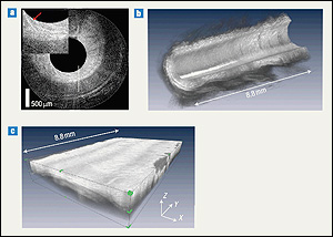
Volumetric renderings of in vivo rabbit colon. a) Single radial frame acquired in 20 ms. Inset shows enlarged view of epithelium, with colonic crypt indicated by a red arrow. b) Cutaway view of the rendered volume. c) Unfolded data set showing the cylindrical volume as a rectangular tissue slab. The entire volume was acquired in 17.7 s.
OCT, developed in the 1990s, provides doctors with micron-scale imaging of tissue in situ and in real time. It enables what Chen refers to as an "optical biopsy"—visualization of changes to tissues without the need for a minor surgery to acquire a sample.
OCT is similar in concept to ultrasound, but the images it produces are created by measuring the echo time delay and intensity of back-reflected light rather than sound. After being inserted into the body by means of a catheter or similar "carrier", an optical beam generated by part of the OCT device—in this case a laser—is scanned across the tissue, and the backscattered light is measured as a function of depth and transverse position to form an image.
Earlier generations of OCT had limited speed that restricted results to two-dimensional images. Thanks to breakthroughs described in the paper, a new generation of devices that use "Fourier-domain OCT" can be 10-100 times faster, and deliver high-resolution, three-dimensional volumetric images. In order to achieve this, Chen, along with colleagues including PI Professor James Fujimoto (MIT), the man who originally developed OCT, combined a fast wavelength-swept laser technology with high-speed data acquisition and signal-processing, and have demonstrated endoscopic imaging capability with high three-dimensional resolution 50 frames per second.
OCT is already in use in the medical community, helping to diagnose diseases. Chen believes the substantial improvements he and his colleagues have made could result in more lives saved. "The purpose [of my group's work] is for early cancer diagnosis in the gastrointestinal tract, especially esophageal cancer, which is a pretty deadly disease," he said. "The 5-year survival rate is only 15%, primarily due to late diagnosis. With a high speed OCT system, we can screen a larger area of the esophagus and select patients with suspicious results for a pinch biopsy. In this way, the chances of missing early cancers can be reduced."
For more information:
Visit Professor Chen's home page »
Published December 19, 2007
Related Stories
Stories / November 20, 2012
Chen Bioimaging Research Wins Prestigious NSF CAREER Award
Stories / January 23, 2012
New Probe Could Help Surgeons Avoid Blood Vessel Damage
Stories / January 19, 2012
New Imaging Technique Covers Middle Ground Between Microscopy...
Stories / November 9, 2010
Kids Learn How Biomedical Imaging Could Save Transplant...
Stories / April 24, 2009
Undergraduates Present Paper at NE BioE Conference

Stories / January 15, 2009
Cancer Detection Research Wins Grant

Stories / Jun 5, 2026
UMD Senior Joshua Mathew Wins 2026 Astronaut Scholarship

Stories / Jun 4, 2026
BIOE Graduate Program Receives Mentoring Excellence Award

Stories / Jun 2, 2026
A “Key” Step Toward Safer Surgeries Worldwide

Stories / May 29, 2026
BIOE Announces Spring 2026 Instructional Impact Awards

Stories / May 28, 2026
Student Spotlight: Outstanding Junior Award Recipient Joel...

Stories / May 26, 2026
Apply: Chair of the Fischell Dept of Bioengineering

Stories / May 15, 2026
BIOE Students Recognized for Innovative Healthcare Solutions at...

Stories / May 13, 2026
2026 Invention of the Year: Engineering Projects Top List

Stories / Apr 28, 2026
Four BIOE Students Named 2026 NSF Graduate Research Fellows

Stories / Apr 20, 2026
To Rid Body of Asthma and COPD, UMD Researchers Hack Into Mucus...
