News Story
Additional Funding for Fluorinated Biomaterials Research
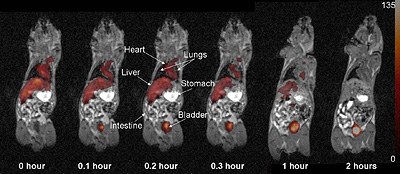
A 19F MRI image from an earlier, similar study showing fluorinated imaging agents passing out of a mouse's system over time.
The parent research study, which is supported by the NIH's National Institute of Biomedical Imaging and BioEngineering (NIBIB), seeks to develop injectable biomaterials that can be used to repair damaged tissue.
"My group pursued the supplemental grant to help speed up the development of peptide-based biomaterials with fluorinated imaging agents embedded in them," Yu, the director of the Drug Delivery and Biomaterials Engineering Laboratory, explains. "We recently made a breakthrough that will give us a significant boost in developing biomaterials that can be monitored non-invasively using fluorine magnetic resonance imaging."
Fluorine magnetic resonance imaging, or 19F MRI, is a type of specialized MRI used to visualize drugs, cells, genes, or implants administered to a patient for therapeutic reasons. "Fluorine MRI is a very useful technology, but its development has lagged far behind that of traditional proton MRI," says Yu. "One of the reasons for this is the lack of a really good imaging agent—what people use now, perfluorocarbons (PFCs), require complex formulation procedures and accumulate in the body, lingering there for months or longer. And because PFCs often emit split 19F signals, the resulting images aren't as clear as they could be. So what we have are huge advances in the field of soft biomaterials, but until now very few in the tools we need to observe how they behave once inside a patient's body."
The Yu Group's discovery centers around the use of molecules called asymmetric bi-spherical fluorinated dendrimers, instead of PFCs. "Because the human body contains no detectable fluorine,19F is perfect for tracking injected or implanted drugs, materials, cells, and genes using MRI—you won't mistake it for anything else." says Yu. "Unlike PFCs, the compounds we developed pass quickly and harmlessly out of the body. They can also serve as sensitive indicators of in vivo biochemical reactions."
Yu believes these qualities will give fluorine MRI the potential to produce much clearer images of implanted materials, which will in turn allow researchers and physicians to better evaluate a biomaterial's interactions with surrounding tissues, including where it is, whether it has degraded, and, if applicable, how much of a drug has been released from it.
Currently, Yu's group is embarking on a new fluorine MRI project that would allow multiple therapeutics to be tracked simultaneously, each with its own color.
For More Information:
Visit Professor Yu's homepage »
See Jiang, Z.-X.; Liu, X.; E.K. Jeong & Yu, Y.B. (2009) "Symmetry-guided Design and Fluorous Synthesis of a Stable and Rapidly Excreted Imaging Tracer for 19F MRI." Angew. Chem. Int. Ed. 48, 4755-4758.
Published October 5, 2009
Recent Stories
Stories / Jun 15, 2026
Alisa Morss Clyne Named Interim Chair of UMD Bioengineering

Stories / Jun 5, 2026
UMD Senior Joshua Mathew Wins 2026 Astronaut Scholarship

Stories / Jun 4, 2026
BIOE Graduate Program Receives Mentoring Excellence Award

Stories / Jun 2, 2026
A “Key” Step Toward Safer Surgeries Worldwide

Stories / May 29, 2026
BIOE Announces Spring 2026 Instructional Impact Awards

Stories / May 28, 2026
Student Spotlight: Outstanding Junior Award Recipient Joel...

Stories / May 26, 2026
Apply: Chair of the Fischell Dept of Bioengineering

Stories / May 15, 2026
BIOE Students Recognized for Innovative Healthcare Solutions at...

Stories / May 13, 2026
2026 Invention of the Year: Engineering Projects Top List

Stories / Apr 28, 2026
Four BIOE Students Named 2026 NSF Graduate Research Fellows
