News Story
Three BIOE Professors Awarded NIH R01s in as Many Months
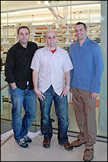
Drs. Scarcelli, Jewell, and Jay
Three University of Maryland Fischell Department of Bioengineering (BIOE) professors each received a National Institutes of Health (NIH) Research Project Grant (R01) between the months of February and April. The awards, which collectively total nearly $5.5 million, will support research in the areas of immunology, optics, and biotherapeutics.
BIOE Associate Professor Christopher Jewell was awarded a four-year, $1.4 million NIH R01 for his efforts to study the immunology behind autoimmune disease and develop new advanced therapies for diseases such as multiple sclerosis.
Autoimmune disease causes the body to identify its own “self” cells as foreign and, in response, the immune system attacks healthy tissue. In MS, the immune system incorrectly recognizes components of the central nervous system, causing inflammation and destruction of myelin, the fatty substance that surrounds and protects nerve fibers. When this happens, nerve fibers and cells are damaged, leading to a loss of motor function and other complications.
To lessen the immune system’s attack against self cells, conventional therapies rely on general immune suppression – a strategy that has proven effective, but can leave patients susceptible to disease or infections.
To explore new pathways toward MS treatments that would leave the patient’s immune system intact, Jewell, collaborator Dr. Jonathan Bromberg (University of Maryland School of Medicine Professor of Surgery and Microbiology and Immunology), and the rest of his team are using degradable biomaterials and combinations of immune signals to study how local changes to the function of lymph nodes – the tissues that determine how immune responses develop – could affect the body’s ability to defend against foreign invaders while inflicting minimal harm to itself. Read More
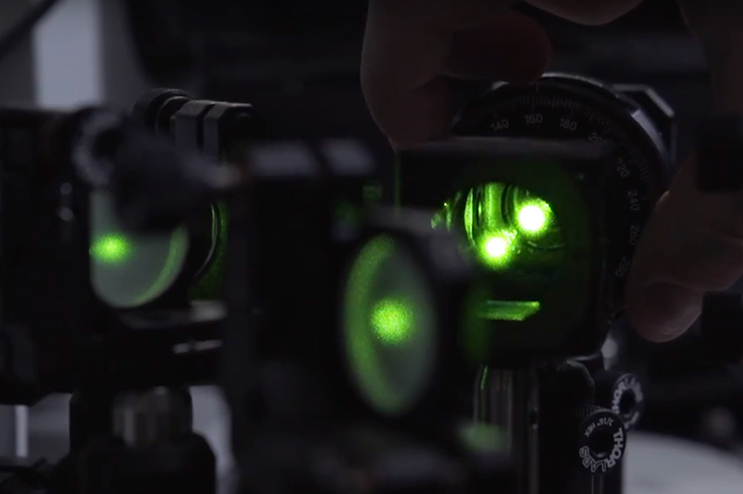
BIOE Assistant Professor Giuliano Scarcelli was awarded a five-year, $2 million NIH R01 for his work towards developing an optical technology that could improve diagnosis and management of keratoconus, a progressive eye disease that often begins during a person’s teens or early 20s.
Keratoconus and related eye diseases are a major cause of vision loss in young adults, the primary concern during refractive surgery screening to treat nearsightedness, and the leading indication for corneal transplants in the United States. Keratoconus causes the cornea – which is normally round – to thin and begin to bulge into a cone-like shape. This cone shape deflects light as it enters the eye on its way to the light-sensitive retina, causing distorted vision.
While it is critical to identify corneal ectasia [weakening of the cornea] as early as possible, current imaging techniques only assess the shape of the cornea and not corneal biomechanics. This leaves doctors and patients with limited information when diagnosing keratoconus, or when planning surgeries.
To overcome current corneal screening limitations, Scarcelli and members of his Optics Biotech Lab have applied their own optical technique, known as Brillouin microscopy, to measure corneal stiffness at a high three-dimensional resolution, without contacting or perturbing the eye.
With support from the NIH, Scarcelli and clinical collaborator Dr. J. Bradley Randleman of the University of Southern California will work to validate Brillouin measurements for mechanical evaluation of the cornea, and characterize observed changes in cases of keratoconus. Moving forward, they also hope to determine the mechanical impact of refractive surgery on the cornea. Read More
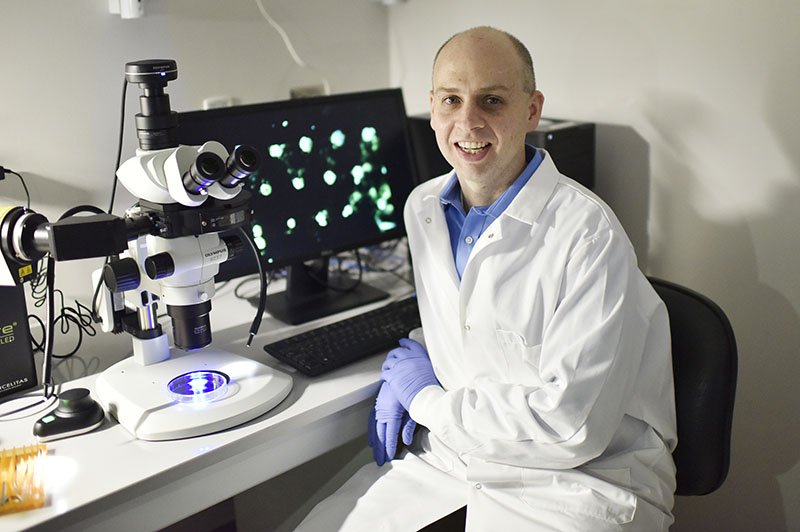
BIOE Assistant Professor Steven Jay was awarded a five-year, $1.9 million NIH R01 award to support his efforts to develop a new approach for treating non-healing wounds using cell-derived structures known as exosomes.
Exosomes are a type of small extracellular vesicle – ubiquitous, biologically-generated structures that naturally transfer nucleic acids between cells. Decades ago, researchers believed that exosomes did little more than offload cellular waste. Today, however, exosomes are better understood as "messengers" that carry out a range of important cellular functions such as the transfer of DNA, RNA, and proteins to other cells, where they are capable of altering the cell’s function. Because of their role in cell-cell communication, exosomes are attractive therapeutic candidates. After all, exosomes are already capable of delivering functional biological cargo to cells – a task that has proven quite challenging to achieve using synthetic systems.
More pertinent to the treatment of non-healing wounds is the role that exosomes can play in mediating a process known as angiogenesis, through which the body creates new blood vessels from preexisting vessels. Angiogenesis is vital to tissue formation and wound healing; a healthy body controls this process using several "on" and "off" switches that either stimulate or inhibit blood vessel formation.
Emerging evidence from numerous research groups around the world shows that exosomes play a crucial role in regulating angiogenesis, with much of the current literature focused on identifying proteins and microRNAs (miRNAs) that contribute to exosome-mediated effects. Jay’s research group has contributed to this body of work with a recent publication in Scientific Reports, identifying a critical regulatory role for specific members of a different class of molecules, long non-coding RNAs (lncRNAs).
Unfortunately, exosomes have a number of shortcomings as potential therapeutic vectors, in particular, they have potentially low potency due to the fact that they contain low amounts of nucleic acid cargo – such as lncRNAs and microRNAs – that are critical to defining their bioactivity.
To overcome this limitation, Jay is working with members of his Biotherapeutic Development and Delivery Laboratory, as well as with Dr. John Harmon, professor of surgery at the Johns Hopkins University School of Medicine, to develop a novel technique to exert control over exosomes derived from endothelial cells – cells that line the interior surface of blood vessels. This approach involves conditioning endothelial cells by exposure to non-toxic concentrations of ethanol, a relatively cheap and readily available chemical compound. This conditioning has the effect of enhancing the expression of specific lncRNAs that promote angiogenesis while diminishing the levels of certain miRNAs that work to reduce angiogenesis. Thus, the overall effect yields production of exosomes with significantly enhanced angiogenic potential that Jay and his team believe could be therapeutically beneficial for wound repair.
Moving forward, Jay and his team will explore how additional exosome production parameters can be integrated with ethanol conditioning to produce highly potent exosomes for wound repair. Read More
Published April 23, 2018
Related Stories
Stories / June 28, 2016
Using Brillouin Microscopy, Scarcelli Aims to Shed Light on How...
Stories / July 15, 2022
Jewell Leads Team in Advancing Multiple Sclerosis Research
Stories / August 16, 2021
Using Light to Attack Cancer

Stories / August 26, 2019
Scarcelli Receives Junior Faculty Outstanding Research Award
Stories / March 11, 2019
New Microscopy Technique Could Change LASIK

Stories / April 20, 2018
Jewell Awarded $1.4 Million NIH Grant for Novel Multiple...

Stories / November 12, 2014
Scarcelli Named NIH K25, Human Frontier Science Program Grant...
Stories / January 5, 2024
Jewell Lab Participates in the Million Mile for Alex's Lemonade...

Stories / March 27, 2023
Joe Huang Recieves Two NIH Funding Awards

Stories / December 20, 2022
UMD Bioengineers’ Brillouin Microscopy Among The Guardian’s...