News Story
Filling the Gaps: Making Better Bone Regeneration a Reality
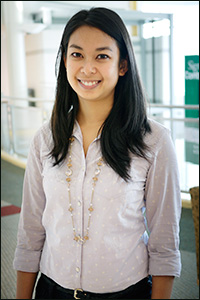
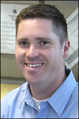
BioE graduate student Bao-Ngoc Nguyen.
Tissue engineering techniques that regenerate bone from the patient's own mesenchymal stem cells seem to be an ideal solution: after differentiating (maturing) into osteoblasts (bone cells), the cells could be implanted in the body in a supportive, biocompatible scaffold. Over time, new bone would grow and fill in breaks, gaps and chips. However, the larger the injury, the more difficult the process becomes.
Clark School Fischell Department of Bioengineering (BioE) graduate student Bao-Ngoc Nguyen (B.S. '11, bioengineering) is part of team out to make large-scale bone regeneration a clinical reality, and her efforts have earned her a 2013 Graduate Research Fellowship from the National Science Foundation (NSF). The three-year award funds her work in the BioE professor and associate chair John Fisher's Tissue Engineering and Biomaterials Laboratory.
The problem with larger injuries, Nguyen says, is ensuring the osteoblasts have an adequate blood supply once implanted. In the lab, the cells are grown in a device Fisher and his group invented, the tubular perfusion system (TPS), which provides them with a supply of oxygen and nutrients. In the body, bone cells on the outside of an implanted scaffold can get the oxygen they need from nearby blood vessels, but by the time those blood vessels grow into the new tissue, it's too late for the cells at the center.
Nguyen contributes to an ongoing effort to co-culture endothelial cells, which grow into blood vessels, with the stem cells so a new vascular network can begin to take root within the developing bone tissue. The hope is that once implanted, the engineered blood vessels will begin to grow and spread, quickly linking up with the surrounding network, bringing vital support to the osteoblasts. Nguyen mimics the body's environment as closely as possible by seeding 3D scaffolds with both kinds of cells and culturing them in the TPS, which has been shown to enhance the growth of engineered tissue over other techniques.
Nguyen says she's excited to be working on the project because of its relevancy and great potential to be translated into real world application. And, she adds, it lays the groundwork for other lifesaving procedures.
"Determining a technique of vascularizing any tissue, not just bone, would be a huge step for tissue engineering applications and allow lab-grown organs to reach a clinical stage," she says. "Because I can see the end result of my research having a tremendous effect on the field of tissue engineering, I am all the more motivated to work hard and reach my research goals."
Nguyen got her start in research as an undergraduate in the bioengineering program, working for former assistant professor Sameer Shah (University of California—San Diego). "I really loved it and learned so much about how to be an effective researcher that I wanted to continue on to grad school," she says. "The [Clark School] has so many opportunities for students, especially because it’s located near many biotech companies and research institutes...I was excited to stay at UMD."
After earning her Ph.D., Nguyen is interested in putting her scientific knowledge to work outside of the lab. Volunteering in the local Vietnamese-American community has given her an appreciation of public policy and advocacy. "I would love to work in the field of science policy," she says. "There is a great need for scientists to help translate and interpret science for law- and policymakers, so they can make more informed decisions."
Published July 7, 2013
