News Story
3D-printed Tissues May Keep Athletes in Action
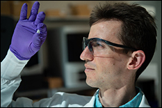
Rice University graduate student Sean Bittner holds a sample of a 3D-printed scaffold that may someday help heal osteochondral injuries of the kind often suffered by athletes. The material mimics the gradient structure of cartilage to bone found at the end of long bones. Photo by Jeff Fitlow
Bioscientists are moving closer to 3D-printed artificial tissues to help heal bone and cartilage typically damaged in sports-related injuries to knees, ankles and elbows.
At the Center for Engineering Complex Tissues (CECT), a National Institutes of Health center at the University of Maryland (UMD), Rice University, and the Wake Forest School of Medicine, scientists reported their first success at engineering scaffolds that replicate the physical characteristics of osteochondral tissue – basically, hard bone beneath a compressible layer of cartilage that appears as the smooth surface on the ends of long bones. UMD Fischell Department of Bioengineering Fischell Family Distinguished Professor and chair John Fisher directs CECT.
Injuries to these bones, from small cracks to pieces that break off, can be painful and often stop athletes’ careers in their tracks. Osteochondral injuries can also lead to disabling arthritis.
The gradient nature of cartilage-into-bone and its porosity have made it difficult to reproduce in the lab, but Rice scientists led by bioengineer Antonios Mikos and graduate student Sean Bittner have used 3D printing to fabricate what they believe will eventually be a suitable material for implantation.
Their results are reported in Acta Biomaterialia.
“Athletes are disproportionately affected by these injuries, but they can affect everybody,” said Bittner, a third-year bioengineering graduate student at Rice, a National Science Foundation fellow and lead author of the paper. “I think this will be a powerful tool to help people with common sports injuries.”
The key is mimicking tissue that turns gradually from cartilage (chondral tissue) at the surface to bone (osteo) underneath. The Biomaterials Lab at Rice printed a scaffold with custom mixtures of a polymer for the former and a ceramic for the latter with imbedded pores that would allow the patient’s own cells and blood vessels to infiltrate the implant, eventually allowing it to become part of the natural bone and cartilage.
“For the most part, the composition will be the same from patient to patient,” Bittner said. “There’s porosity included so vasculature can grow in from the native bone. We don’t have to fabricate the blood vessels ourselves.”
The future of the project will involve figuring out how to print an osteochondral implant that perfectly fits the patient and allows the porous implant to grow into and knit with the bone and cartilage.
Mikos said the collaboration is a great early success for CECT, which works to develop bioprinting tools to address basic scientific questions and translate new knowledge into clinical practice.
“In that context, what we’ve done here is impactful and may lead to new regenerative medicine solutions,” Mikos said.
Co-authors of the paper are Rice graduate student Brandon Smith, postdoctoral researcher Luis Diaz-Gomez, undergraduate Carrigan Hudgins, Anthony Melchiorri, University of Maryland Fischell Department of Bioengineering alum and associate director of the Biomaterials Lab at Rice, and David Scott, the Noah Harding Professor of Statistics; and John Fisher, CECT director and Fischell Family Distinguished Professor and chair of the University of Maryland’s Fischell Department of Bioengineering. Mikos is the Louis Calder Professor of Bioengineering and a professor of chemical and biomolecular engineering, of chemistry and of materials science and nanoengineering.
The National Institutes of Health and the RegenMed Development Organization supported the research.
Published April 1, 2019
Related Stories
Stories / August 11, 2020
Fisher Elected IAMBE Fellow

Stories / October 1, 2019
4D Bioprinting Smart Constructs for the Heart
Stories / January 4, 2018
Fisher Named Tissue Engineering Co-Editor-in-Chief

Stories / June 23, 2017
Fisher, Bracaglia Weigh in on the Future of Regenerative...
Stories / August 20, 2014
Bracaglia, Melchiorri Awarded American Heart Association...
Stories / June 25, 2026
Grand Challenges 2.0: Engineers Partner to Unlock Solutions

Stories / February 26, 2026
AIM Welcomes 5 Distinguished Artificial Intelligence Scholars

Stories / February 9, 2026
Struggling With Fibroids, UMD Researcher Seeks to Engineer a...

Stories / February 6, 2026
UMD Bioengineering Contributes to ARPA-H PRINT Program Focusing...

Stories / January 30, 2026
BIOE Researchers Publish Study Advancing Probiotic Vesicle...
