News Story
BIOE Capstone Class Presents 20 Novel Projects
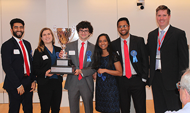
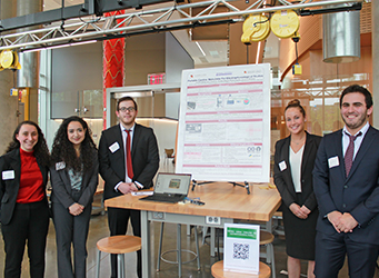
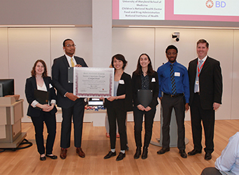
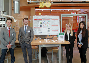
 Showcasing the many ways bioengineers are working to improve human health, the Fischell Department of Bioengineering (BIOE) 2019 Senior Capstone class exhibited a total of 20 novel concepts during the Capstone II finale on May 13, 2019.
Projects ranged from a system for rapid toxin removal to a brain-to-brain synchrony detector for improved psychotherapy.
Ninety-six BIOE students pitched their products to a panel of esteemed judges, BIOE faculty and other students. The annual Capstone Design Competition has become a marquee event, capping off the academic year. It was created and is sponsored by Susan Fischell, wife of A. James Clark benefactor Dr. Robert E. Fischell.
This year's panel of judges featured:
-
University of Maryland, Baltimore collaborators
The Fischell Department of Bioengineering receives support for its Capstone Design competition from the Fischell family, the Mpowering the State initiative, Dr. Ivor T. & Ms. Michelle Knight, and industrial sponsors AgNovos and Becton, Dickinson and Company. Additionally, Capstone students often receive mentorship support from industry representatives and/or clinicians, including investigators from the University of Maryland School of Medicine, Children's National Health Center, the U.S. Food and Drug Administration (FDA), and the National Institutes of Health (NIH).
2019 Capstone Teams, Projects, and Awards:
 Team 1: ElutoCAT Drug-Eluting Thoracic Catheter
Emily Blick, Christopher Hiner, Katherine Jones, Drishti Maniar, and Thea Ornstein
Advisors: Christopher Jewell (BIOE Associate Professor and Associate Chair) and Dr. Joseph S. Friedberg (University of Maryland School of Medicine)
BEST POSTER
Thoracic surgery is an operative process that focuses on chest organs such as the heart, lungs, esophagus and the trachea. The surgery results in significant postoperative and chronic pain which is mainly caused by the insertion and continuous placement of a thoracic catheter in the patient’s pleural space for up to 2 weeks. The insertion of the thoracic catheter, or better known as the chest tube, is imperative to drain air, fluid and blood post-operation. Current pain management strategies such as local analgesic injections, thoracic epidural or intravenous painkillers, fail to deliver localized, sustained pain relief to patients for the entirety of the time period in which they have a chest tube placement. Thus, Team 1 aimed to provide long-term patient pain relief by designing a silicon, drug-eluting thoracic catheter which would slowly elute lidocaine at the site of chest tube insertion, over a one week period. To demonstrate proof of concept, the research group experimented on small slabs of silicon catheter. The optimal dissolvable polymer, solvent and polymer to lidocaine ratios were determined via the dip-coating method. By performing UV spectroscopy and scanning electron microscopy, Team 1 demonstrated that they effectively coated a portion of a silicon chest tube in a polymer-lidocaine mixture and successfully eluted lidocaine from the coated tube into phosphate buffer saline over an hour. Future work will focus on increasing the amount of lidocaine loaded in order to reach physiologically relevant levels as well as key manufacturing processes that will enable product scale-up.
 Team 2: Knee Extension Monitoring Device for Children with Cerebral Palsy
Viswanath Gorti, Zain Kazi, Nikita Kedia, Mateo Reveiz, and Janette Yacynyc
Advisors: Advisors: Dr. White (BIOE Associate Professor and Associate Chair) and Dr. Pergami (Children’s National)
FIRST PLACE
Cerebral palsy (CP) is a neurological disorder which impairs movement, muscle tone, and posture. Clinical studies have found differences associated with knee angles in children with CP relative to healthy children that result in difficulty walking. Unlike healthy children, children with CP are unable to extend their knees to a full 180° when walking, resulting in a crouched gait pattern. These patients must be monitored by clinicians, including physical therapists, to ensure proper knee extension. This treatment can be time consuming and expensive. According to the Center for Disease Control and Prevention, the lifetime cost for a person with CP is 1.175 billion dollars, including physician visits, assistive devices, therapies, rehabilitation, and other long term-care options. The average copay for a weekly physical therapist appointment is $30 and patients with CP would need constant monitoring of this movement, which would accrue additional costs. Existing knee angle monitoring devices on the market are expensive and not tailored for pediatric patients. Here, Team 2 presented a low-cost device that will allow patients to receive at-home, direct feedback based on the knee angle measurements. The patient will be notified with the percentage of steps he or she took correctly and the gait cycles will be automatically detected and classified via a software application. This recorded data can also be stored and sent to the physician for daily tracking of the patient’s progress. Two approaches were compared for measuring knee angles: a stretchable strain sensor that modeled a parallel plate capacitor and resulted in a voltage drop with stretch, and a linear taper potentiometer in which the analog signal was converted to the corresponding angle. After testing both sensors, it was determined that the potentiometer system provided more precise and reliable angle measurements. Knee angle measurements for healthy gait and crouched gait were recorded and compared. In the future, Team 2 will pair their device with a step sensor to allow for a more detailed analysis of the patient’s gait. The group plans on testing their device clinically and this testing will be validated against the current physical therapy treatment.
 Team 3: Minimally Invasive Glucose Sensing Device
Medina Algoba, Kyran Gibson, Mike Mistretta, Alex Paskal, and Cynthia Uzoukwa
Advisors: Dr. Edwards Eisenstein (BIOE Associate Professor) and Dr. Johana Diaz (University of Maryland School of Medicine)
According to the World Health Organization, an estimated 15 billion infants are born preterm every year. Preterm birth complications are the leading cause of death in children under the age of 5. Infants are considered preterm when born earlier than thirty-seven weeks. These infants are at a high risk of severe health complications requiring them to be constantly monitored. Premature infants are kept in isolettes that control their environment where they are connected to a multitude of machines that monitor their vitals constantly. Continuous heath monitoring allows nurses and doctors to determine the best treatment each infant needs to facilitate growth. One of the most common blood tests is the blood glucose test. Blood for this test is collected via a heel prick, an arterial line, or venipuncture.
 Team 4: A Laparoscopic Tool for Pediatric Epicardial Lead Removal
Lauren Fowlkes, Amelia Hurley-Novanty, Manav Parikh, Shaanit Sen, and Alex Wahl
Advisors: Dr. Hubert Montas (BIOE Associate Professor) and Dr. Rohan Kumthekar (Children’s National)
Infants with severe cardiac problems may require implantation of a pacemaker into the abdomen with a lead which is attached to the epicardial surface. These pacemakers only last up to 10 years, and eventually must be replaced with a transvenous lead. During this procedure, the epicardial lead, which lies in the abdominal and thoracic cavities, is abandoned except in emergency situations. Given that these leads have adhesions to vital organs and directly to the heart, removal requires open heart surgery. Leaving this lead results in decreased quality of life for these patients since they can no longer undergo magnetic resonance imaging procedures or go through metal detectors without issue. Current laparoscopic devices, which would make the procedure less invasive, are not appropriately sized for a pediatric population. Additionally, many tools are difficult to use and require many manual inputs to achieve relatively little mobility. Team 4 has designed and prototyped a laparoscopic device with five degrees of freedom and higher precision than current models, in order to remove adhesions in the size scale found in pediatric patients. This device couples existing electrocautery components with a novel articulation mechanism, which includes an articulating tip directed by mechanical surgeon inputs. The articulation mechanism within the body has a small,
 Team 5: High-Flux Hemofiltration System for Rapid Toxin Removal
Morgan Hoffman, Daniel Hopkins, Morgan Janes, Milan Patel, and Manaahil Rao
Advisors: Dr. Helim Aranda-Espinoza (BIOE Associate Professor and Associate Chair), Dr. Alison Grazioli (University of Maryland Medical Center), Dr. Zhongjun Wu (University of Maryland School of Medicine), and Dr. Josh King (University of Maryland School of Medicine)
SECOND PLACE
Acute poisoning is a clinical problem that affects 2.16 million people each year. Hemofilters, which are designed to remove fluid from the blood during cardiac bypass, can also be used to filter toxins from the bloodstream in certain toxic ingestions. Unfortunately, current hemofilters lack the filtering capacity for many traditionally non-dialyzable toxins, resulting in limited clinical applicability for critical ingestion patients. Commercially available hemofilters lack adequate surface area and are unable to filter blood at high enough rates to remove toxins with a large volume of distribution or high protein binding fraction. Extracorporeal membrane oxygenation (ECMO) is an adjacent technology designed to treat heart and lung failure that circulates blood at high flow rates (4-5 L/min). While ECMO is unable to provide toxin removal, in recent years it has been increasingly used to offer hemodynamic support for critically ill patients suffering from toxic overdose. The goal of this project was to develop an ECMO-compatible hemofiltration system to facilitate rapid and efficient acute toxin removal at high flow rates. By increasing the overall surface area and incorporating the system directly into an ECMO circuit, Team 5 sought to overcome traditional challenges with hemofilters to enable rapid filtration of traditionally non-dialyzable toxins. Team 5 developed a prototype toxin filtration system by creating a parallel “circuit” of eight connected hemofilters. This system can support an input flow rate of 4 L/min, and it was able to remove the non-dialyzable toxin flecainide from saline at an improved and clinically relevant rate. The team's final results show a 16-fold increase in the removal rate, with potential for a 21-fold increase, in comparison to traditional hemofiltration. The clinical contribution of this project is a robust, time-saving toxin removal modality for patients who have overdosed on traditionally non-dialyzable drugs.
 Team 6: MR Surgery: Mixed Reality Protestate Surgery from Patient-Derived MRI Scans
Christian Haryanto, Anjana Hevaganinge, Hannah Horng, and Madeleine Noonan-Shueh
Advisors: Dr. Lan Ma (BIOE Lecturer), Dr. Minhaj Siddiqui (University of Maryland School of Medicine), Dr. Axel Krieger (University of Maryland)
Prostate cancer affects millions of Americans, and often requires a radical prostatectomy to remove both the prostate and the tumor to treat the cancer. Following the surgery, an anastomosis must be performed to replace the bladder and the urethra. Surgeons often have difficulty identifying where the prostate and surrounding anatomy are, and must take extreme caution not to damage other organs, including the neurovascular bundle. Team 6's goal is to leverage recent developments in mixed reality to create surgical guidance and training data for surgeons to more effectively complete surgery. Their models use virtual reality to simulate the mechanical stresses that cause deformation of the prostate and other organs during surgery. This data can be used to generate data for augmented reality simulations that allow automated tracking and labeling of the prostate, as well as an interactive display of a patient-derived 3D model of the prostate. The team's simulations can reduce the cost of this vital surgery and increase the quality of patient care by providing surgical guidance during prostatectomy.
 Team 7: Portable Cardiac Stimulator for Electrophysiological Studies
Andrew Amick, Larry Feldman, Shireen Khayat, Paula Kleyman, and Amanda Marques
Advisors: Xiaoming (Shawn) Ming (BIOE Professor) and Rafael James (Children’s National)
Heart function can be characterized and analyzed using electrophysiology (EP), a branch of biology that pertains to the propagation of ion current in the heart. EP studies utilize cardiac stimulators, a device that outputs programmed electrical pulses, to initialize and study impulse propagation through different areas of the heart by inducing arrhythmias. When combined with quantification or visualization of the changing electric potential in the heart, EP studies provide insight on disease pathologies. EP studies can also be used to assess the effects of different substances on heart function, including environmental toxins and newly developed pharmaceuticals. Current cardiac stimulators are very large, bulky devices that are difficult to move between rooms and high cost, making it undesirable, but necessary to have to purchase a separate device for each room. Due to the significant impact these studies have, there is a need to increase the usability and portability of the cardiac stimulator while making them affordable and efficient. To do so, we have created a prototype of a portable external cardiac stimulator that mirrors and improves the functionality of the cardiac stimulator currently used at Children’s National Hospital by utilizing a microcontroller as a more cost effective and intuitive alternative. Team 7's device is compacted to allow for quick transport, thereby eliminating the need and costs associated with having multiple devices. It maintains the basic, most used functions of current cardiac stimulators, allowing user regulation of different pulse parameters and offering an option for synchronized pulsing that utilizes real-time ECG data to determine ideal pulse intervals. The device includes user interface software usable on any personal computer that regulates a microprocessor controlled electrical output. The output is designed to interface with a stimulus isolator that isolates, modifies, and delivers the precise signal through leads that attach to the heart. The microprocessor is also able to read in the pulses it outputs, as well as the surface electrocardiogram signals provided by an amplifier. It then can send the values to the interface for real time plotting. The team's portable cardiac stimulator will make EP studies more accessible and less laborious for researchers and clinicians.
 Team 8: Noise-Reducing Electromagnetic Latch System for NICU Isolettes
Jillian Carter, Austin Goncz, Sara Merlock, Samuel Mircoff, and Blair Smith
Advisors: Dr. Yu Chen (BIOE Associate Professor) and Dr. Rose Viscardi (University of Maryland Medical Center)
Recent research has indicated that decibel levels above 45 dB can impact normal neurological development in premature neonates. It was discovered that this level of noise, and more, is created in the Neonatal Intensive Care Unit (NICU) by the monitoring instrumentation, human activity, and the opening and closing of portholes on isolettes. To mitigate the potential effects of ambient noise, NICU staff now separate infants into individual, quieter rooms. However, the excessive level of noise created by the opening and closing of the current porthole latch mechanism still remains a problem. Team 8 has designed a new electromagnetic latch system that will allow the NICU staff to gain immediate access to the child in case of an emergency, while generating less noise than current latch mechanisms. The new mechanism is controlled via a foot pedal, proximity sensor, and Arduino Uno microcontroller. The proximity sensor detects when the porthole door is nearly closed and activates the electromagnet, locking the door shut while still allowing manual access in case of emergency. The foot pedal then provides a direct, hands-free mechanism to turn off the electromagnetic, and open the isolette door quickly. The electromagnet and proximity sensor are retrofitted onto the current isolette design via a custom 3D printed housing unit that attaches in place of the original latch. This new mechanism decreased max noise output while opening and closing the doors by over 20 dB, but did not lower it to the goal of below 45 dB. This could be due to ambient noise in the NICU, which ranges from 50-55 dB at baseline. Further work for this design includes making the circuit more robust and providing a finish that would be more suitable for sterilization.
 Team 9:Erythrocyte Mediated Angiography Verification with Phantom Eye
Danielle Firer, Matthew Kim, Rachel Luthcke, Richard Silcott, and Drew Vogel
Advisors: Dr. Giuliano Scarcelli (BIOE Assistant Professor) and Dr. Osamah Saeedi (University of Maryland Medical Center)
Recent studies have shown that impaired ocular blood flow is implicated in the progression of Glaucoma, Diabetic Retinopathy, and Age-Related Macular Degeneration, three of the leading causes of blindness worldwide. To facilitate early detection of these pathologies, researchers have developed techniques for measuring ocular blood flow. One of the primary methods is erythrocyte mediated angiography (EMA). When performing EMA, blood is extracted from the patient and mixed with Indocyanine Green (ICG) to label the red blood cells by fluorescence. The ICG-labeled blood sample is reintroduced into the patient’s body and a Heidelberg Retinal Tomograph (HRT) is used to capture an angiogram, where the ICGlabeled blood cells appear as brighter pixels. These cells are used to measure blood cell speed and track ocular blood flow in all patients. When imaging with HRT, the patient’s native cornea is used for additional magnification; however, corneal dimensions and vessel position in the eye vary across all people. This study investigated the assumption that axial distance (AD) of the blood vessel and/or the focal length (FL) of the lens does not affect HRT images. Three FL (15mm, 18mm, 20mm) and three AD (18mm, 20mm, 22mm) were tested. For all trials, blood flow was fixed at a rate of 0.005mL/min and cell speed of 10610.33µm/s. Only trials collected at an AD of 22mm were within one standard deviation of the expected speed; however, the 15mm lens could not focus on the vessel at 22mm. Image resolution increased as AD increased, but decreased as lens FL increased. Team 9's device was able to successfully investigate the impact of lens strength and axial distance on HRT measurements by comparing incident red blood cell speed. The application of this system has the potential to accelerate research related to the development of more accurate diagnosis and monitoring of major causes of blindness worldwide.
 Team 10: SpiCast - Safe Transportation for Pediatric Patients with Femur Fractures
Deven Appel, Danya Chowdhury, Shweta Roy, and Richard Sellars
Advisors: Dr. Lex Schultheis (BIOE Research Professor) and Dr. Matthew Oetgen (Children’s National)
STUDENTS' CHOICE AWARD
Pediatric femur fractures are the second most common long bone fracture in kids. Nineteen out of every hundred-thousand children in the United States experience femur fractures every year. Within the past year, an estimated 14,600 children between the ages of 0 to 11 had a femur fracture. These fractures can be as a result of birth, hip dysplasia, or due to unintentional falls and accidental injuries. Treatment varies and depends heavily on the child’s age. However, the common form of treatment for children under the age of five is immediate spica casting, which is an orthopedic cast that immobilizes the hip or thigh. Everyday car seats do not provide the space, comfort or ease for spica cast patients. Although spica casts are the best method of treating children with femur fractures, they are extremely inconvenient, especially when it comes to transporting these children from place to place. Team 10 has created an attachment that can be inserted into the regular car seat of the patient, to be inexpensive, feasible, and comfortable. The prototype was created using an assortment of materials, including a vacuum-sealable bag, expandable foam, and fiberglass. The final product consists of a foam attachment that is tailored to the child and their respective car seat. After finalizing the design, a drop test was performed with the insert attached to a car seat to evaluate the impact of a fall on the child and car seat. From these results, Team 10 found that the acceleration on the baby was reduced by 2 m/s2 with the carseat, however, the acceleration drop remained consistent between a 3 ft and 28 ft drop with our setup. Overall, the proposed prototype offers several societal benefits such as reduced parental strain, reduced financial burden, and improved safety and comfort for injured children. Future work can be conducted that includes material exploration, consumer testing, and NHTSA confirmation.
 Team 11: GaitMate: A Gait Monitoring Solution for Pediatric Cerebral Palsy
Tolulope Awosika, Nicole Duster, Jenny Katsnelson, Stacey Mannuel, David Tucker
Advisors: Dr. Li-Qun Zhang (BIOE Professor), Dr. Lan Ma (BIOE Lecturer), Dr. Paola Pergami (Children’s National), and Dr. Kevin Cleary (Children’s National)
BEST ABSTRACT
Cerebral palsy affects three out of every 1,000 children in the United States. With spastic cerebral palsy comprising the majority of cases, many of these children suffer from abnormal gait patterns such as toe-walking which lead to future health issues such as stiffened leg muscles. While the current standard of care involves patients attending monthly physical therapy sessions, the relative infrequency of these visits often leads to relapses and therefore minimal progress. To facilitate for more frequent physical therapy for substantial gait improvement, a pressure-monitoring shoe sole has been developed. Four piezoelectric pressure sensors are attached to the top of a sole at strategic points which allow for one to distinguish between gait patterns. The sensors are then hooked up to an Arduino and Bluetooth to transmit data wirelessly to either a laptop or mobile application. After fabricating and testing the prototype, it has been concluded that the device is capable of tracking and recording an individual’s gait in real time. It can detect both heel-toe and toe-heel walking and display distinct patterns. The response time of the device is rapid enough to accommodate a normal walking pace and is responsive to a wide range of applied weights. Further testing with a range of pediatric patients will be the next step in finalizing this product to provide the most accurate and comfortable product. Once on the market, the affordability and ease of use of this product will allow for patients to easily conduct physical therapy on a daily basis, increasing the likeliness of improved gait performance.
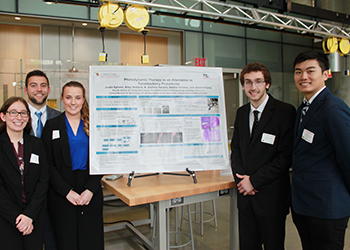 Team 12: Photodynamic Therapy as an Alternative to Tonsillectomy Procedures
Jason Chiang, Natalia Ochman, N. Zachary Rausch, Justin Sylvers, and Arley Wolfand
Advisors: Dr. Huang Chiao Huang (BIOE Assistant Professor), Dr. Kevin Cleary (Children’s National), Dr. Avinash Eranki (Children’s National), and Dr. Rahul Shah (Children’s National)
A tonsillectomy is one of the most common surgical procedures in the United States, with more than 530,000 performed annually as of 2011. In this procedure tonsils are removed due to recurrent inflammation, sleep disorders, or complications from being enlarged. The procedure can be performed by different modes of excision such as cold knife surgery and electrocautery; however, complications of all tonsillectomies include primary and secondary hemorrhages, infections, and a long recovery. The goal of this project was to validate an alternative procedure for tonsil removal using photodynamic therapy (PDT). The envisioned procedure involves topical application of a photosensitizing drug (PS) to the tonsil followed by a wait interval allowing the drug to localize. After this, the tonsil is to be irradiated with a low-powered diode laser causing the excitation of the PS, generation of cytotoxic species, and cell death. If the PS can localize in and destroy germinal centers of the tonsil, the tonsils should postoperatively shrink away after several weeks without cutting and the associated bleeding of standard excision.
The specific aims of the work conducted for this project were to 1) characterize the penetration of liposomal benzoporphyrin derivative (L-BPD), an FDA approved PS, into excised tonsil tissue after topical application in solution, and 2) characterize the ability of irradiation following L-BPD application to cause cell death in excised tonsil tissue. To this end, both penetration and cell death studies were performed. For the drug penetration studies, fluorescence microscopy was used to measure penetration after application of L-BPD in varying concentrations and with varying wait intervals. The greatest penetration was observed with 70 µM L-BPD and a 30-minute incubation. For cell death studies, tonsil portions were treated with L-BPD, irradiation, both, or neither. While evidence of necrosis was visible in all groups, it was qualitatively most pronounced in the groups that had both the drug and laser applied.
These results suggest that L-BPD is capable of penetrating excised tonsil tissue and, in combination with irradiation, induces cell death. This project is a promising preliminary step toward validating PDT for tonsil removal; however, further work needs to be done to validate the procedure.
 Team 13: Orthopedic Surgical Training Model for AGN1 Local Osteo-enhancement Procedure (LOEP)
Navid Chowdhury, Daniel Lambert, Jingce Lei, Rhea Puhtumana, and Josephine Stair
Advisors: Dr. Angela Jones (BIOE Senior Lecturer) and Dr. Jonathan Shaul (AgNovos Healthcare)
Osteoporosis is a metabolic disease that reduces the density and quality of bone. As the bone becomes more porous and fragile, there is an increased chance that a fracture may occur. AgNovos has developed a new synthetic biomaterial called AGN1 to replace and restore functionality to osteoporotic bone. In the local osteo-enhancement procedure (LOEP), ANG1 is injected into the femoral head of the patient. Currently, AgNovos employs a customized femur model made by Sawbones as a training model for simulation and training. The purpose of this project is to create an alternative training model for the LOEP surgery that is cheaper, more realistic, and more practical to use. Team 13's model has three principle components: a reusable outer case, disposable femur head and flesh inserts, and a tutorial application. The 3D-printed outer case simulates the upper thigh and holds the bone model in place. The femur head was created by setting polyurethane foam in a silicon mold and coating the shaped foam in epoxy resin. The optimal mixing ratio of the polyurethane foam was determined through a nail-weight impact test. The flesh insert is comprised of Humimic Medical Gel and a thin layer of silicon, simulating muscle and skin, respectively. The application provides an intuitive walk-through of the training procedure using our model. Team 13 met their goal of developing a cheaper alternative to the $70 Sawbones model. The total one-time cost of our case was $53.35, and the cost for a set of disposable femur head and muscle inserts is $12.07. The team's femur model is watertight, allowing trainees to perform the irrigation and aspiration steps of the LOEP procedure, and the case itself allows the bone to be easily inserted and replaced during training. Overall, the group has created a suitable alternative to the currently-used Sawbones model that is cheaper, more realistic, and easier to use.
 Team 14: PID Controlled Hemofiltration for Poison Control
Vaidehi Bhagat, James Bookhultz, Elliot Bromberg, Akshaya Ganesh, and Chris Kuffner
Advisors: Dr. Alison Grazioli (University of Maryland Medical Center), Dr. Josh King (University of Maryland Medical Center), Dr. Zhongjun Wu (University of Maryland Medical Center)
According to the U.S. Renal Data System Annual Report, there are currently over 660,000 Americans being treated for kidney failure and 468,000 of those patients are on dialysis. The kidney has several functions, including filtering blood and the excretion of wastes and toxins from the body. When a patient has renal failure they often undergo dialysis, a process in which the patient is attached to a machine which performs the filtering functions of the kidney, mechanically. Although dialysis is typically used for patients undergoing chronic renal failure, and often doesn’t handle sufficient enough blood volumes to treat acute renal failure. The Maryland Poison Control center handles about 44,000 calls a year regarding people who have been poisoned with substances such as household chemicals, snake venom, alcohol or opioids.
Hemofiltration will be used to treat patients suffering from acute renal failure rather than traditional dialysis. High hemofiltration clearance levels can be achieved by taking advantage of Extracorporeal Membrane Oxygenation (ECMO). ECMO technology is capable of oxygenating the body’s blood volume at a rate of 5L/min. By applying this technology to hemofiltration, thousands of lives could be saved.
Proportional Integral Derivative (PID) control was implemented using an Arduino microcontroller in order to set the flow rate for fluid replacement equivalent to the flow rate of effluent being filtered out from the system once the saline solution from the main centrifugal pump was filtered. The user could set the flow rate at which the effluent was being filtered out and the system would correct the flow rate to that value. Results of the testing conducted showed that the relationship between Lpm and read voltage was approximately 0.1V for every 0.2 Lpm of flow. This relationship was needed to convert the desired setpoint to a voltage that could be compared to the voltage being read by the flow sensor. Based off of the information received by the sensor, the controller can adjust the flow rate as necessary. The PID controller was tested at two different flow rates against a PI controller to determine which overshot less at the two flow rates. It was found that the PI controller was favorable in both cases of flow rates. The PI controller resulted in less overshooting and and a faster response in the case of the 0.5 Lpm trial. The controller was observed to cause more overshooting at higher flow rates. This device will improve outcomes for patients with acute renal failure cases that could not be treated using current methods.
 Team 15: Scapholunate Ligament Augmentation System for Rapid Functional Restoration
Alec Boyle, Chris Damiani, McKenzie Figur, and Adriana Meriles-Medina
Advisors: Dr. Keith Herold (BIOE Associate Professor) and Dr. R. Frank Henn III (University of Maryland Medical Center)
The scapholunate ligament serves a critical role in stabilizing the wrist and providing support during physical activities. However, its prominent role and complexity within the carpal system make ruptures of the scapholunate ligament (SLL) one of the most common and complicated types of wrist injuries. Multitudes of methods exist to surgically repair the system to regain functionality, but these methods are inadequate, and many have long-term side effects such as high re-tear rates, reduced range of motion and flexibility, and an inability for the patient to return to peak performance. The current procedures are highly dependent on the stage of the scapholunate dissociation (SLD). The six stages are categorized by quantifying the degree of tear (complete or partial), the possibility of repair of the ligament, alignment or malalignment of the scaphoid, and cartilage health. Team 15's device will be applicable to complete tears of the ligament with and without malalignment of the scaphoid, which correspond to stages 2 through 4. The team proposed to work towards solving this medical problem with applications of mechanics and surgical engineering principles. Under the guidance of clinical mentor, Dr. R. Frank Henn III, an orthopedic surgeon at the University of Maryland School of Medicine, the team designed a reliable method for surgically augmenting the ruptured scapholunate ligament to restore functional mobility without sacrificing long-term stability. The group's intent was to establish a method of repair that is common across orthopedic practices and replaces the multitude of less desirable methods used today. The purpose of the team's device is to augment the scapholunate ligament as it heals for stage 2 SLD or to replace the native ligament for stages 3 and 4 SLD. Their design consists of two spacers made of polypropylene that can be inserted into the scaphoid and lunate. Much like drywall anchors, these spacers expand with the insertion of polyether ether ketone (PEEK) screws and are held in tension with polyethylene suture. The maximum force the native SLL ligament can withstand is 260 N. Both FEA simulations and mechanical testing have shown that the group's device is capable of holding far greater loads than required, with a safety factor above 2. The team's device provides the load-bearing capabilities to effectively hold the scaphoid and luminate in tension while providing the flexibility necessary to return to functional movement quickly.
 Team 16: Vesicular Delivery of Interleukin-11 Gene for Osteoporosis Treatment
Elizabeth Bentley, Corinne Farley, Matt Mulvaney, Maria Pozo, and Eric Wang
Advisors: Steven Jay (BIOE Assistant Professor) and Dr. Stephen Thom (University of Maryland, Baltimore)
MPOWER AWARD
Osteoporosis is a disease characterized by a reduction in bone density, resulting from an imbalance in the rates of bone deposition and bone resorption. Current treatments for osteoporosis include various medications, such as hormone-related therapy, that often put the individual at risk for blood clots, heart disease and several cancers. To address these challenges, gene therapy utilizing extracellular vesicles (EVs) could be used to promote osteoblastogenesis. Due to their self-origin, flexibility, and ability for surface conjugation, EVs mitigate immunogenicity and systemic clearance of other drug delivery systems. Here, we “transfect” EVs with plasmid DNA and carry genetic content to target cells, facilitating gene expression. Specifically, Team 16 delivered a model fluorescence plasmid to human embryonic kidney cells and saw observable expression of GFP. The team’s results indicate the feasibility of patient-derived EVs as personalized plasmid delivery systems.
 Team 17: In-Line Blood Analyzer for Premature Infants
Sarah Asfari, James Fookes, Adam Herskovits, Devon Parsons, and Cristina Tous
Advisors: Dr. Silvina Matysiak (BIOE Associate Professor) and Dr. Sripriya Sundararajan (University of Maryland School of Medicine)
THIRD PLACE
The health and development of a premature infant in the neonatal intensive care unit (NICU) is currently assessed by measuring the concentration of various analytes at any given time. The typical analytes that are measured include: lactate, oxygen saturation, glucose, urea, creatinine, sodium, potassium, magnesium, and calcium. Blood testing is currently used to determine the concentration of these analytes. These blood tests require a new 5 milliliter sample to be taken each time the concentration of an analyte is to be determined. The accumulation of these blood draws during the early postnatal period can result in the infant losing an estimated 10-20 mL of blood/kg per week, or 15%-30% of their circulating blood volume in the first weeks of life. This loss in blood volume commonly results in the premature infants becoming anemic, and also places a large amount of stress on their aerobic, skeletal, and cardiovascular systems. Team 17 eliminated the need for blood withdrawal by designing an in-line blood analyzer that determines analyte concentration using Fourier Transform Infrared Spectroscopy (FTIR). The analyzer consists of a catheter line from the premature infant attached to one input of a flow through cuvette. The other input of the cuvette contains a port for a syringe to be attached. The blood can be drawn into the cuvette by applying suction via a syringe. The flow through cuvette can then be placed into an opaque box containing the FTIR spectrophotometer, which will measure the amount of light absorbed by the blood sample. The concentration of the blood analytes are then determined by inputting the absorption values into Beer’s law. The concentration of specific analytes are targeted by using absorption values of peaks at specific wavelengths that are known to be characteristic of a molecule. Once the concentration is determined, the blood can be returned back into the infant’s body by pushing down on the plunger of the syringe. Due to cost restraints, an FTIR spectrometer could not be obtained. An infrared Arduino photodiode was used as a placeholder for the FTIR spectrometer to demonstrate proof of concept. The specific peaks for glucose were determined to occur at wavenumbers of 1012 cm-1 and 1081 cm-1, and isolated by subtracting out the spectrums of other analytes found in blood. Additionally, the extinction coefficient to be used in Beer’s law to determine the concentration of glucose was calculated to be 0.023 ,and the path length of L·mm mmol our design was found to be 4mm. Overall, Team 17 has succeeded in designing the framework for a novel way to measure the concentration of various analytes in the blood of a premature infant that does not require them to experience any loss in blood volume. The analyzer can be improved upon to accomplish the entire objective of detecting the concentration of all analytes by installing the proper FTIR sensor, and isolating the appropriate peaks, as was done for glucose.
 Team 18: Single-Stage Percutaneous Tracheostomy Device
Subhashini Arumugam, Zon Fatima, Daniel Najafali, Gabriel Sanz, and Alya Suraya
Advisors: Dr. John P. Fisher (BIOE Department Chair) and Dr. Joseph Rabin (University of Maryland)
ADVISORY BOARD AWARD FOR TRANSLATIONAL DESIGN
A percutaneous tracheostomy is a bedside-procedure that circumvents airway obstructions and offers ventilatory support to patients via a deliberate opening in the trachea. After dilating an initial incision, a tracheostomy tube is placed in the opening to maintain airflow. Current devices used for dilation require multiple rounds of instrumentation leading to increased risk of bleeding, infection, and tracheal damage during the procedure. Further limitations in the device structure can result in a lack of passive airflow, leading to hypoxia. Thus, Team 18 proposed a single-stage percutaneous tracheostomy device that further reduces this range of complications, decreases the rounds of instrumentation needed to place the tube, and improves upon tactile feedback during the procedure. A passive airflow component to address the risk of hypoxia during a tracheostomy will also be included. A prototype of the group’s device, including a novel dilation mechanism, was created using 3D printing technologies, exhibiting enhanced structural detail and cost-efficiency when compared to other methods. Considerations of the biocompatibility as well as structural integrity of our device has been investigated and refined in this initial design. Team 18 demonstrated efficacy of their model by testing on 3D-printed models of the trachea. The group aims to conduct further testing to assess overall procedure time, ventilation, and incidence of misplacement. Upon successful execution, such a design will not only minimize patient complications, but also procedure costs and operating burden on the physicians, making the procedure more accessible and improving patient outcomes.
 Team 19: Improved Performance of N95 Isolation Masks: Enhancing Communication, Fit, and Comfort
Nicole Cavett, Athenia Jones, Julian Kopelove, Kailey Mihavetz, and Priscilla Seah
Advisors: Dr. William Bentley (BIOE Professor) and Dr. Jeffrey Hasday (University of Maryland Medical Center)
Numerous studies show that healthcare workers are poorly compliant with respiratory protection guidelines, especially when a N95 respirator is recommended. Many features of the current N95 masks may contribute to the lack of compliance, such as temperature and humidity accumulation in the mask, as well as the fact that pressure from the nose clip and elastic straps may cause discomfort when the N95 is worn for prolonged periods of time. To improve compliance and to reduce the risk of disease transmission, Team 19 developed a prototype with features that improve the fit and comfort of the mask. One-way exhalation valves and novel filter media were used to reduce the temperature and humidity accumulation. Viscoelastic polyurethane foam was used to not only generate a better seal between the mask and the user’s face, but also to reduce contact pressure. To facilitate communication, Team 19 has incorporated a clear polyethylene terephthalate (PET) centerpiece, which permits lip reading and facial expressions to be seen. Preliminary data suggests the prototype increases humidity within the mask while decreasing temperature. Future work includes improving the fit of the mask to greater variety head forms. In addition, Team 19 plans to continue development and testing of the filter media, while also testing effectiveness of a desiccant. Ultimately, the group hopes to accomplish the intended social impact of improving compliance and reducing the risk of disease transmission.
 Team 20: Detection of Brain-to-Brain Synchrony for Improved Psychotherapy
Michael Dib, Akash Gami, Idrisa Rahman, and Leah Ross
Advisors: Yang Tao (BIOE Professor), Dr. Zhi-De Deng, (National Institute of Mental Health), Dr. Marc Lener (Singula Institute)
Current models to treat mental disorders mainly include cognitive behavioral therapy (CBT), however, a main limitation of CBT is that it offers no way to determine or measure the therapeutic alliance between the clinician and the patient. Previous studies have shown that for two people interacting with each other, there is a neural association that develops between them. This idea can have great implications in the field of psychotherapy. The detection of brain synchrony between a patient and a therapist can show how engaged they are with each other during a therapy session. Although psychotherapy is an established field and treatment for mental disorder alleviation, technologies that allow the accurate detection of a large array of biologically generated signals are relatively new and constantly being renovated. The most advanced technology used currently for standard protocol during clinician-patient interaction in therapy sessions is simply a voice recorder. Devices and methods such as electroencephalography (EEG), facial pattern recognition, and electrodermal detection are used almost exclusively for scientific research. The implementation of these methods into psychiatry and other mental health-related fields has limitless potential in improving the efficacy of clinician-patient interactions. While it would traditionally take several sessions for a clinician to understand the individual needs of a new patient and to decide the most appropriate methods of therapy, these technologies could give the clinician a far greater idea of how to approach the patient even by the second session. The product Team 20 developed aims to capitalize upon the concepts of social interaction and hyperscanning to be able to detect and measure the therapeutic alliance between the clinician and the patient by detecting brain to brain synchrony signals via EEG. This provides the greatest ecological validity due to its non-invasiveness and its ability to be used in a typical social face-to-face therapist-patient interaction.
Published May 17, 2019
Related Stories
Stories / September 12, 2022
UMD Bioengineering Advances in Rankings
Stories / May 9, 2022
BIOE Capstone Competition Highlights 24 Novel Projects

Stories / May 21, 2020
BIOE Capstone Class Virtually Presents 24 Novel Projects

Stories / May 15, 2025
From the Chesapeake Bay to Deep Space: Innovating for the...

Stories / December 4, 2024
UMD iGEM Team Wins Gold Medal at 2024 Paris iGEM Jamboree for...
Stories / June 4, 2024
Meet the First Cohort of the MARC Program
Stories / May 31, 2024
BIOE Students Showcase Award-Winning Innovation at Capstone Expo
Stories / May 8, 2024
MARC Program Now Accepting Applications
Stories / May 8, 2024
A Ventilation Coach for Opioid Overdose Bystanders Takes Top...
Stories / May 2, 2024
Bioengineering Celebrates Undergraduate Excellence