News Story
BIOE Researchers Draw New Connections between Brain Environment, Breast Cancer Metastasis

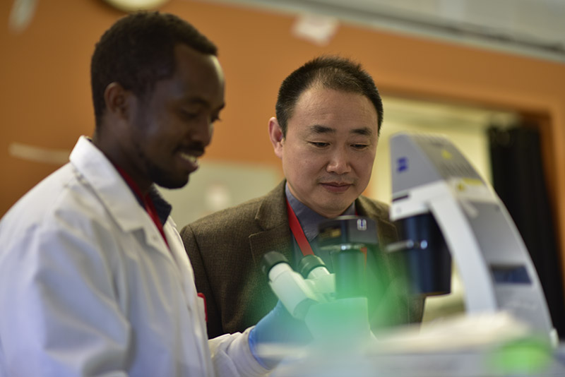
Researchers from the University of Maryland Fischell Department of Bioengineering (BIOE) have discovered a connection between biochemical cues from cells in the brain environment and breast tumor cell migration. Their findings, published in “Astrocytes from the brain microenvironment alter migration and morphology of metastatic breast cancer cells” in the FASEB Journal, point to key factors that cause breast cancer to metastasize to the brain.
“Metastasis to the brain is one of the most deadly aspects of breast cancer, and it often leads to a poor prognosis for patients,” said BIOE assistant professor and alumna Kimberly Stroka (Ph.D. ’11). “When tumor cells travel from the breast to the brain, they get transported through the bloodstream. Once they enter the brain, they need to travel out of the bloodstream into the brain environment, commonly referred to as brain tissue. But, there is still so little researchers know about how and why tumor cells cross what is known as the blood-brain barrier to enter this space.”
The blood-brain barrier (BBB) is a semi-permeable barrier made up of brain endothelial cells that line the blood vessels in the brain. This barrier plays a critical role in protecting the brain from foreign substances in the blood, while allowing essential nutrients to reach the brain. Unfortunately, the BBB is so effective that it also hinders the delivery of potentially life-saving therapeutics to the brain.
To understand why breast tumor cells can overcome this barrier – while many targeted treatments cannot – Stroka and researchers from her Cell and Microenvironment Engineering Lab looked to study astrocytes, one of the most abundant cell types found in the brain. Astrocytes play a key role in supporting the BBB and brain homeostasis, but recent research indicates that they are also linked to brain metastasis and tumor cell survival across the BBB. In fact, Stroka and her team have found that biochemical cues from astrocytes actually increase the speed of breast tumor cell migration by changing their shape.
Even more, the research group discovered that when they applied the same biochemical cues directly to the extracellular matrix – the collection of proteins and carbohydrates that surrounds the cells – there was an even greater increase in tumor cell velocity.
"These findings are significant because they show that biochemical cues from astrocytes could be affecting tumor cell behavior directly or through modifications to their extracellular matrix,” said BIOE graduate student Marina Shumakovich, first author on the paper. “Understanding these mechanisms, such as examining the role of astrocyte-secreted matrix metalloproteinases – enzymes that degrade the extracellular matrix and are secreted by astrocytes – brings us closer to understanding cancer metastasis to the brain and thus potentially developing new targets for therapeutics."
Moving forward, Stroka and her lab group aim to further explore how biochemical cues from astrocytes can change the extracellular matrix in such a way that impacts tumor cell migration. One way they hope to do this is by developing a BBB-on-a-chip model that will allow them to accurately mimic the brain endothelial layer such that they can use engineering strategies to inform development of regenerative therapies for diseases.
###
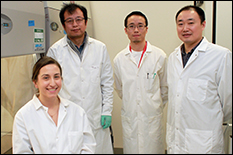
Researchers from the university’s Fischell Department of Bioengineering (BIOE) and National Institutes of Health (NIH) National Heart, Lung, and Blood Institute Laboratory of Developmental Neurobiology contributed to the FASEB Journal paper. The full list of authors is: first author Marina A. Shumakovich (BIOE), Caitlin Mencio (NIH), Jonathan Siglin (BIOE), Rebecca Moriarty (BIOE), Herbert Geller (NIH), and Kimberly Stroka (BIOE, NIH, UMB).
The research is supported by a Burroughs Wellcome Career Award at the Scientific Interface.
Published October 16, 2017
Related Stories
Stories / July 20, 2022
Combating Metastatic Cancer Through Blood Draw
Stories / October 17, 2019
Stroka Named 2019 Young Innovator of Cellular & Molecular...
Stories / January 19, 2021
Waging War on Metastatic Cancer
Stories / July 7, 2020
Huang Receives NIH Trailblazer Award & NSF CBET Grant

Stories / June 25, 2019
Python-Based Program Could Advance Understanding of Blood-Brain...

Stories / February 25, 2019
Study Outlines Targeted Treatment Option for Aggressive Breast...
Stories / November 17, 2017
Stroka Named Outstanding Young Scientist by the Maryland...

Stories / June 29, 2017
New Microscopy Technique Sheds Light on How Cells Sense...

Stories / June 12, 2017
John Schardt Awarded American Association of Pharmaceutical...

Stories / June 5, 2017
Muro, IBBR Faculty Awarded Maryland Innovation Initiative Grants