News Story
Chen Bioimaging Research Wins Prestigious NSF CAREER Award
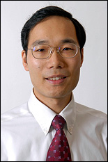
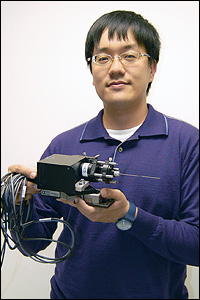
Fischell Department of Bioengineering assistant professor Yu Chen.
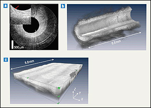
Chen specializes in the advancement of optical coherence tomography (OCT), a micron-scale imaging technology that allows doctors to examine changes to tissues in the body and in real time, without the need for a biopsy to acquire a sample. OCT is similar in concept to ultrasound, but creates images by measuring the echo time delay and intensity of back-reflected light rather than sound.
Chen's award-winning project, "Multi-Scale Imaging of Neural Interconnects Using MRI-compatible Parallel Photonic Needle Interface," will apply his group's innovations to the design of a new system capable of observing neural activity in the brain. The system, which will use OCT and magnetic resonance imaging (MRI) in parallel, is expected to improve our understanding of fast neuronal activation and the slower changes in blood flow that occur in response.
"Functional MRI is particularly useful for visualizing the location of neural activity, but has limited spatial resolution and temporal response—about one millimeter and one second, respectively," explains Chen. "Optical techniques such as OCT offer high spatial and temporal resolutions—about one to ten microns and one to ten milliseconds, respectively. A system that combines and correlates large field-of-view MRI and high-resolution optical imaging can enhance our understanding of brain function and improve the accuracy of image-guided neurosurgery."
The new system is based in part on the Needle OCT/Doppler (DOCT) probe, a long, slender bioimaging device Chen's Biophotonic Imaging Laboratory designed for neurosurgeons. The needle imaging probe, which measures less than 1mm in outside diameter, is capable of accompanying surgical tools into the brain inside a tube called a cannula, looking ahead and interpreting the biological landscape for the surgeon, who can then make course corrections that avoid damaging blood vessels while en route to a tumor or injury.
Chen is excited about the project's potential. "The MRI/parallel optical imaging platform will enable multi-scale imaging of neural structure and function deep within the living brain for the first time," he says. "It has the potential to link the microscopic neurovascular functions discovered in animal models with the macroscopic signals relevant for human imaging, thereby enabling translation of basic biomedical research insights to clinical applications."
The NSF CAREER program supports the career development of outstanding junior faculty who most effectively integrate research and education within the goals and missions of their programs, departments, and schools. Chen is the fifth BioE assistant professor since the department's launch in July 2006 to receive a CAREER Award, preceded by Ian White earlier this year, Joonil Seog (now with the Department of Materials Science and Engineering) in 2011, Adam Hsieh in 2009, and Helim Aranda-Espinoza in 2007.
Published November 20, 2012
